Inguinal Canal: Anatomy and Important Clinical Conditions
Are you in search of anatomical details related to the inguinal canal? You are at the right point to learn the precise details relevant to the inguinal region and associated inguinal canal.
Considering human anatomy, there are multiple regions and openings that provide a passage for different other structures. These openings are referred to as foramina, hiatus, or canals. Specifically, inguinal canals are located in the inguinal region. The inguinal region has specific boundaries extending from the lower abdomen to the middle region of the upper thighs of every side.
These are paired structures in the lower region of the anterior abdominal wall. In addition, the anterior abdominal wall lies in a craniocaudal position from the xiphisternum. Moreover, it limits near the lower border of the lower eight ribs up to the inguinal ligaments and pubis.
What are the anatomical landmarks of the inguinal canal?
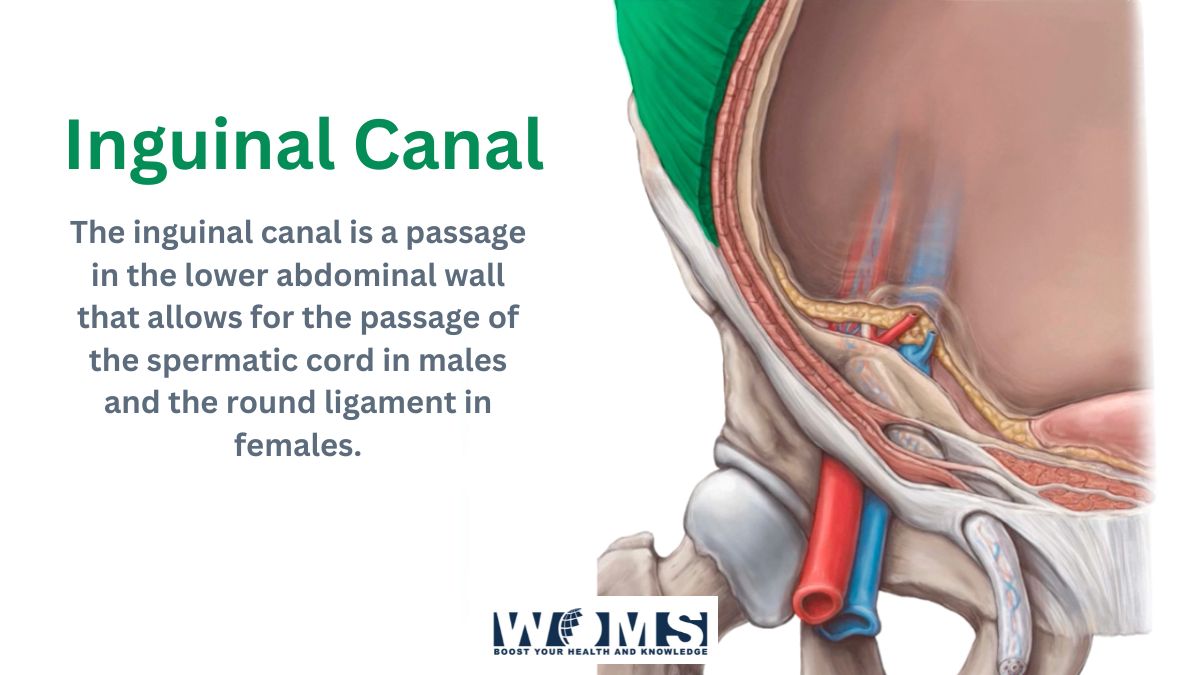
The inguinal canal is an obliquely arranged intramuscular slit of about 3 to 5 cm in length. Superolaterally, it originates from the deep inguinal ring. It runs along the midpoint of the inguinal ligament between the anterior superior iliac spine and pubic tubercle. In addition, the deep inguinal ring can be located at a level of 1.25 cm above the mid-inguinal point. It terminates at the level of the superficial inguinal ring.
The superficial inguinal ring is located 1 cm superolateral to the pubic tubercle.
What are the boundaries or associated walls of the inguinal canals?
They lie in accordance with different other structures. These structures are the boundaries of the canal. Through the identification of these structures, a surgeon can easily get access to the inguinal canals. For this reason, it is important to locate the boundaries of the canals. Here is detailed information related to the limiting structures of the canals.
Anterior side – the anterior wall or side contains skin, superficial fascia, aponeurosis of external oblique muscles, and a smaller part of the internal oblique muscle. These structures lie from outer to inner regions.
Posterior side – the posterior surface contains the conjoint tendon medially, insertion area of external and internal oblique muscles, and transversalis fascia quite laterally.
Superior side or roof – the roof contains muscles. It is formed by the internal oblique and transversus abdominis muscles.
Inferior wall or floor – the floor is formed by multiple ligaments, including inguinal and lacunar ligaments. This portion consists of the medial aspect of these ligaments.
You may also like to go through this detailed article on Essential structures of Portal Triad.
What are the contents of the inguinal canal?
The content of the inguinal canal varies from gender to gender. In males, there are different structures as compared to females. The ilioinguinal nerve, lymph nodes, and blood vessels are common in both sexes.
Let us discuss the content details of the canal according to gender.
Contents in females
In females, there is a round ligament of the uterus that passes through the inguinal canal. This round ligament reaches the posterior aspect of the labia majora to insert.
Contents in males
The male inguinal canal mainly contains the spermatic cord. Besides this, the spermatic cord contains layerings of fascia, multiple arteries, nerves, and other reproductive structures. There are multiple structures present in the spermatic cord, but ultimately residing in the canal.
- Fascia is the continuing layers of the muscular and fascial layers. There are three types of fascia surrounding the spermatic cord.
- The deepest of these fascias is the internal spermatic fascia. It is the continuation of transversalis fascia.
- The middle layer is the cremasteric fascia. It is the continuation of the internal oblique muscle and fascia.
- The outermost layer of the spermatic cord is the external spermatic fascia which is the continuation of the external oblique aponeurosis.
- There are three main arteries including the arteries to the vas deferens, the testicular artery, and the cremasteric artery.
- There are three major anatomical structures that are pampiniform plexus, vas deferens, and testicular lymphatics.
- There are three common nerves that include the genital branch of the genitofemoral nerve, sympathetic and visceral afferent fibers, and the ilioinguinal nerve.
The most important clinical point related to the inguinal canals is the inguinal hernia. An inguinal hernia is the pathological protrusion of the viscera beyond the walls of the containing cavity. It may cause weakness in the walls. Inguinal hernia can be direct or indirect.
- Direct hernia can be due to protrusion of the abdominal viscera with the help of the posterior wall. It occurs mainly during adulthood.
- Indirect hernia occurs due to the deep inguinal ring. It is the commonest type and includes 50% of the population. In addition, it occurs mainly on the right side and is prone to be congenital.
- Inguinal endometriosis is also a clinical aspect related to the canal.
- Neoplastic growth may also develop in any part of the canal.
- There may be varicosities of the round ligament in the females and varicocele in the males.
Conclusion
Inguinal canal serves as an opening or passage in the anterior abdominal wall. It is located at the medial half of the inguinal ligament. There are different structures of the inguinal canal comparable to both sexes. But, the ilioinguinal nerve is common to both sexes. The inguinal canal is also important because of its clinical relevance in the form of an inguinal hernia.
Frequently asked questions (FAQs)
What is the physiologic role of the inguinal canal?
The inguinal canal serves the main role to provide support to the oblique muscles, abdomen, and pelvic region. In addition, it provides flexibility to the pubic area and supports the soft tissues in the groin region.
What is the rule of 3 for the inguinal canal?
The inguinal canal contains multiple structures. For ease, there is a rule of 3 to avoid the complexity of the structural arrangements. There are three fascial layers, three arteries, and three nerves.
What is the other name of the inguinal canal in males?
The other name for the inguinal canal in males is referred to as the gonadal artery. Inguinal terms usually explain the area of the groin or the lowest lateral aspects of the abdomen.