The ultimate guide to Dog Skull: 3 types and Images
There are a variety of dogs with different shapes and sizes of dog skull. Some are more popular, and some are more expensive than others. But what is most important is what your dog means to you and your relationship with them. Your dog skull is a unique and special part of your furry friend’s life.
This article will discuss some anatomical features and types of the dog skull. Let’s get straight to it.
Types of Dog Skull
They are of three types, each with its characteristics. The following types are discussed in detail.
- Mesaticephalic Skull
- Dolichocephalic Skull
- Brachycephalic Skull
Mesaticephalic Skull
Another name is mesocephalic, and the shape of the skull is square. The dogs having mesaticephalic heads have skulls of the same length and width. This breed has broader snouts and nasal cavities, enhancing its scenting capabilities.
The well-known breeds with Mesaticephalic skulls include Chihuahua, Labrador, Retriever, and Beagle.
Dolichocephalic Skull
The literal meaning of Dolichocephalic means ‘Long Skulled’. The dogs with dolichocephalic skulls have longer craniums and shorter cranial widths. In this type of skull, a long and narrow snout enables a great range of vision. The long snout makes sniffing, hunting, and tracking easier for them and as a result, makes them a perfect breed among other dogs. The Dolichocephalic skull is considered one of the long skulls.
The breeds having Dolichocephalic skull includes Whippet, Greyhound, and Saluki
Brachycephalic Skull
This skull type is fascinating with its cute appearance and short skull. The shorter snouts do not let the warm air cool down.
Brachycephalic breeds are English Bulldog, Lhasa Apso, and Bullmastiff.
Shape of Dog Skull
A dog’s skull is determined by the shape of the animal’s brain. The skull has to be shaped in such a way that it can accommodate the size and weight of an animal’s brain. A dog skull is different from a human’s skull because dogs have much smaller brains than humans. The shape of a dog skull is an essential feature that differentiates different breeds.
The shape of the skull can be classified into three categories: round, long and flat. A round skull is rounded on both sides, a long skull is longer than wide, and a flat skull has little or no curvature. The shape of the dog’s head depends on its breed. Different breeds have different forms because they are bred to do specific tasks such as hunting, herding, or guarding livestock.
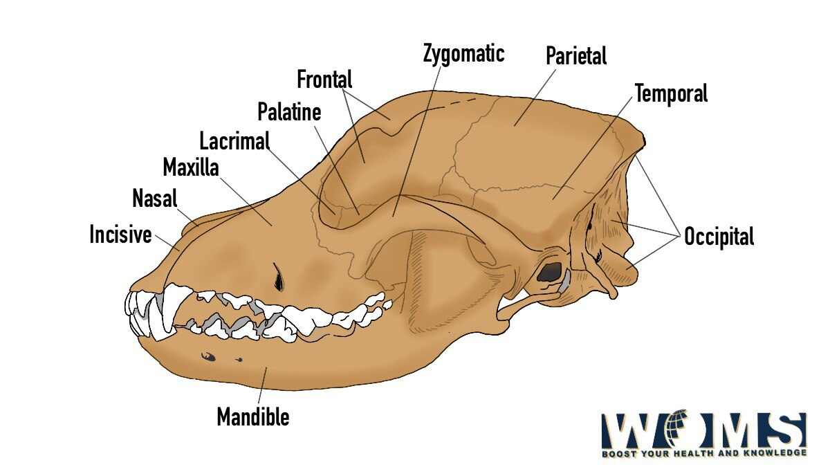
Bone Anatomy of Dog Skull
The skull of dogs come in many shapes and sizes but share the same basic anatomy. The dog skull is made up of several bones that are fused together. The shape of the skull varies depending on the breed of dog. The bones in a dog skull include:
- Occipital Bone
- Interparietal and Parietal Bone
- Frontal Bone
- Temporal Bone
- Sphenoid Bone
- Ethmoid Bone
- Maxilla Bone
- Palatine Bone
- Pterygoid Bone
- Zygomatic Bone
- Mandible
Let’s discuss each in detail to understand the dog skull anatomy
Occipital Bone
The occipital bone consists of two parts: the basilar and the squamous part. Before birth, the squamous part joins with the interparietal bone. The basilar part fuses with the tympanic bulla. A mastoid foramen is present at the junction of the squamous part of the temporal bone. This foramen is located dorsal and rostral to the foramen magnum.
An angular nuchal crest is present caudally in a dog. You will find two tubercles located ventrally to the nuchal crest.
Look for the following structures while identifying the occipital bone of the dog skull:
- Basilar and Squamous part of the occipital bone
- A nuchal crest and tubercles of occipital bone
- A mastoid foramen of dog skull
Parietal and Interparietal bone
As discussed above, the squamous part of the occipital bone joins with the interparietal bone resulting in the formation of the interparietal process. The interparietal bone also helps in the formation of the transverse sinus.
You can identify the parietal bone by its rhomboid shape and extreme curve. Parietal bone plays a significant role in forming the roof of the cranial cavity of the dog.
A sagittal crest can be seen at the junction between the right and left parietal bones. The external surface of the parietal bone makes the temporal fossa.
While identifying the dog skull, you need to focus on the following structures:
- Sagittal crest of the dog skull
- Temporal fossa of the skull
- External and Internal parts of the dog
Frontal Bone
The frontal bone is irregularly shaped, with some osteological characteristics. The frontal bone consists of an orbital part, a frontal squama, a temporal surface, and the nasal part. You will notice a short zygomatic process in the dog skull. There is a foramen known as supraorbital foramen on both sides of the supraorbital. An ethmoidal foramen is present in the frontal bone.
You need to search these parts to identify the dog skull.
- Short zygomatic process
- Supraorbital foramen
- Temporal line
Temporal Bone
Temporal bone plays a vital role in making the ventrolateral wall of the calvaria. The temporal bone contains the following parts:
- Petrosal
- Squamous
- Tympanic
A cerebellar fossa is present at the caudomedial side of the petrosal of the temporal bone. Internal acoustic meatus is at the ventral side of the cerebellar fossa.
The tympanic part is located ventrally, and it is more prominent by its structure which is known as bulla tympanic.
The squamous part has a long and curved process known as the zygomatic process. A mandibular fossa is located ventrally to the zygomatic process.
Sphenoid Bone
The sphenoid bone occupies two-thirds of the cranial cavity in a dog skull. The two structures, presphenoid and basisphenoid, are present in the sphenoid bone. The basisphenoid takes part in the formation of the middle cranial fossa. A pterygoid process is located in the basisphenoid bone.
Ethmoid Bone
The ethmoid bone is between the facial and cranial parts of the dog’s skull. The following structures make the ethmoid bone:
- Two ethmoid labyrinths
- Median perpendicular plates
- A cribriform plate
The ethmoid labyrinths take part in the formation of the bulk of the ethmoid bone.
Maxilla Bone
In dog skull anatomy, it is a short bone located caudally. Maxilla has a body and four processes. The external surface contains a prominent structure known as the infraorbital foramen. The medial surface has a nasal part that has numerous crests.
Palatine Bone
Palatine bone is located caudomedial to the maxilla bone in the dog skull. Palatine has two laminae; horizontal and perpendicular laminae. The horizontal laminae make one-third of the hard plate.
A nasal crest is present at the nasal surface of the palatine bone, which joins with the vomer bone.
The perpendicular laminae help in making the nasopharyngeal meatus.
Pterygoid Bone
In dog skull anatomy, the pterygoid bone is small, thin, curved, and has four-sided plate bone. A pterygoid hamulus is located at the caudoventral angle of the pterygoid bone. The pterygoid fossa is present on the medial surface of the pterygoid bone.
Zygomatic Bone
In dog skull anatomy, the zygomatic bone forms the rostral half of the zygomatic arch. The Zygomatic bone is curved and long. There are two surfaces and two processes in the zygomatic bone.
The lateral surface of the zygomatic bone is convex and located transversely. The orbital surface of the zygomatic bone is concave and medial to the bone.
Mandible
You will notice six alveoli for incisor teeth and two for the canine teeth in the dog’s mandible. A short interalveolar space can be seen in the body of the dog’s mandible. The rostral part of the mandible extends medially and contains two or three mental foramina.
You need to observe these structures while identifying the dog’s mandible.
- Mandibular notch
- Masseteric fossa
- Coronoid process
- Condyloid process
FAQs
Are dogs’ skulls thicker than humans?
it is true to some extent that a dog’s skull is thicker and stronger than a human’s. The medium and large-sized dogs’ skulls are thicker due to the muscle mass, which is more than that of humans. Contrary to that, small dogs’ skulls are not thick.
Do dogs’ skulls change shape?
Yes, it does change. The change in the shape of the dog skull occurs due to the morphological changes in the brain.
How do I identify a dog skull?
Dog skull identification is not easy, but one can do this by knowing all the anatomical features and types of the dog’s skull.
Why does a dog’s skull have a ridge?
The ridge is a sign that the dog has strong jaw muscles. The chewing muscle, temporalis, is attached to the sagittal crest. The development of the sagittal crest is associated with the development of the temporalis muscle.
What does a dog skull look like?
The dog skull has various shapes, and it entirely depends on the breed to decide whether it is dolichocephalic or mesaticephalic.