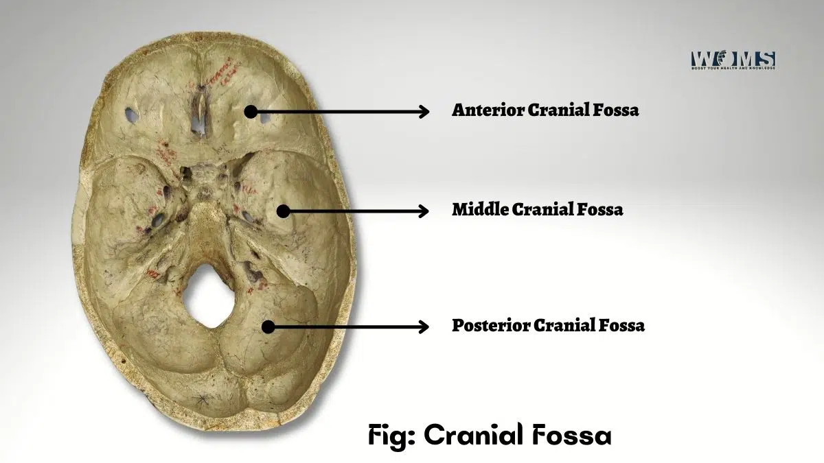
Cranial Fossa: Contents, Anatomy and Clinical Significance
The cranial fossa is the skull cavity that contains the brain and most vital structures, including the eyes, nose, mouth, ears, and sinuses. The base of the skull (occiput) is formed by the occipital bone at the back of the head, which encloses the foramen magnum, a large opening in the center of the skull through which the spinal cord passes. The parietal bones form the sides of the skull; these bones meet across the top of the head to form the frontal bone.
The front part of the skull comprises two pieces: the frontal and the nasal bones. At the bottom of the skull, the temporal bone forms the roof of the cranial fossa. It has three parts: the superior, middle, and inferior portions. The upper portion of the temporal bone includes the external auditory meatus or ear canal, the tympanic membrane, and the eustachian tube. The lower portion includes the mastoid process, which is shaped like an inverted cone and is covered with skin. On its outer surface lies the oval window, a small hole that allows sound waves to pass from the outside world into the inner ear.
Anterior cranial fossa
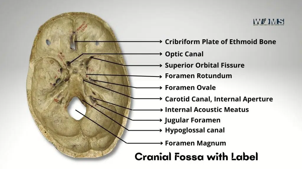
The anterior cranial fossa is bounded by the frontal bone, the ethmoid bone, and the sphenoid bone. The floor of this space is formed by the frontal bone, while the ethmoid bone forms the roof. The walls of the fossa consist of the maxillary bone on one side and the palatine bone on the other. This area of the skull is where the eye sockets are located. The orbit is the hollow space within the skull that surrounds the eyeball.
The orbits are divided into four compartments: the lateral, medial, superomedial, and inferolateral compartments. Each compartment is separated by bony partitions called optic canals. These canals allow nerves to pass from the eye to the brain. The optic nerve travels down the optic canal of the lateral wall of the orbit.
Middle Cranial Fossa
This skull area is bounded by the cerebrum’s frontal lobe of the cerebrum, the parietal lobe of the cerebrums, and the occipital bone. The floor of the fossa is formed by the parietal bone, while the roof of the fossa is composed of the squama of the occipital bone and the temporal bone.
The walls of this area are made up of the frontal bone, the zygomatic arch, and the pterygoid plates. The lateral wall of the fossa is lined by the temporalis muscle, which covers the temporomandibular joint. The middle cranial fossa is home to the pituitary gland, which produces growth and thyroid-stimulating hormones.
Posterior Cranial Fossa
Located behind the posterior end of the skull, the posterior cranial fossa is surrounded by the occipital condyles, the quadrigeminal cistern, and the cerebellar tonsils. The floor of the posterior cranial fossae is formed by the occipital bone, while the roof consists of the cerebellum.
The walls of the posterior cranial fossa are formed by the tentorium cerebelli, which separates it from the cerebral hemisphere. The fourth ventricle is located immediately below the cerebellum. It is filled with cerebrospinal fluid produced by the choroid plexus. The cerebellum controls balance and coordination.
Anatomy
The bones of the human skull form an enclosed cavity known as the cranial vault. They protect the delicate brain tissue from injury. The cranial vault comprises 12 pairs of bones, including the frontal, parietal, temporal, and occipital bones.
The bones of the skull have several functions. For example, they protect the brain and spinal cord. They also support facial structures, such as the eyes, nose, mouth, teeth, ears, and jaw. The skull protects the brain from injuries caused by blows or falls. The bones of the cranial vault also help hold the brain in place.
Skull Base
The skull base is the junction of the bony elements of the skull. It includes the maxilla, mandible, sphenoid, ethmoid, palatine, and basisphenoid. The skull base is divided into four parts: the anterior part, the middle part, the posterior part, and the basilar part. A suture separates each part.
The nasal septum and the orbit bound the anterior part of the skull base. The middle part of the skull base lies between the optic canal and the otic capsule. The skull base’s posterior part extends from the skull’s midline to the foramen magnum. The basilar part connects the occipital bone to the first cervical vertebrae. The skull base’s basilar part forms the cranial cavity’s posterior wall.
Function
The cranial fossae are spaces within the skull that protect various brain parts. They also house structures that regulate body temperature.
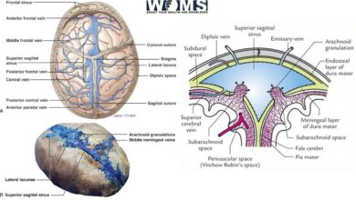
Frontal sinuses
These are small cavities between the frontal bones and the frontonasal suture. There are six pairs of these sinuses, but only five are visible externally. They are named according to their location: frontal, orbital, lacrimal, maxillary, and sphenoidal sinuses.
The frontal sinuses are located on top of the frontal bone. The largest sinuses are the frontal sinus, which extends from the nasal cavity to the inferior margin of the orbit. The frontal sinuses protect the brain from infections by allowing air to flow freely through them. The frontal sinuses have openings called Ostia. When an infection occurs, pus can collect in the frontal sinuses and then be drained out through the Ostia. The frontal sinuses also help keep the nose cool because they act as a chimney when air passes through them.
Orbit
This is the space surrounding the eye. It is divided into two compartments: anterior and posterior. The anterior compartment is separated from the posterior compartment by the optic nerve. The posterior compartment is further subdivided into the infraorbital canal and the ethmoidal canal.
The orbit is bordered laterally by the periorbital fat pads. Medially, the orbit is bound by the zygomatic process of the frontal bone. Posteriorly, the orbit is bounded by the inferior orbital rim. The supraorbital notch forms the medial boundary of the orbit. The lateral boundaries of the orbit are formed by the zygomatic processes of the frontal bone.
Optic Nerve
This is the bundle of nerves that carries visual information from the retina to the thalamus and other brain areas. The optic nerve begins at the back of the eyeball, where the retina is attached. It runs forward along the side of the head, passing through the optic canal.
In the middle of the optic nerve is the optic disc, the point at which light enters the eye. At this point, the nerve fibers divide into two bundles, one going to the left side of the brain and another to the right. These bundles travel together until they reach the optic tract, which is part of the optic radiation. From there, the fibers split again, sending some to the temporal lobe and others to the parietal lobe.
Ethmoidal Canal
This is a narrow passage leading from the nasopharynx to the olfactory region. It is lined with mucous membrane and contains lymphoid tissue. This area is important in the immune system as it provides a site where antigens enter the bloodstream.
Nasal Cavity
The nasal cavity consists of three regions: the superior, middle, and inferior meatus. The superior meatus is the opening of the nostril. The middle meatus is the passageway connecting the superior and inferior meatus. The inferior meatus is the opening between the middle and lower turbinate’s. The nasal cavity has several functions, including respiration, speech, and respiratory tract protection. The nasal cavity is filled with soft tissues such as connective tissue, cartilage, muscles, and glands.
Turbinate
These are conical projections extending from the sides of the nasal cavity. Each turbinate is covered by a thin layer of skin known as the mucosa. Turbinates comprise loose connective tissue, bone, cartilage, and muscle. The function of the turbinates is not fully understood. Some believe that they play a role in warming or humidifying inhaled air.
Nerve
There are many different types of nerves within the body. They can be classified according to their location, cell type, size, or function. There are cranial nerves (in the skull), spinal nerves (within the spine), sympathetic nerves (from the thoracic ganglia), sensory nerves (sending messages to the central nervous system), and motor nerves (carrying impulses away from the central nervous system).
Sympathetic Nervous System
The sympathetic nervous system controls involuntary actions such as heart rate, blood pressure, sweating, and digestion. The sympathetic nervous system originates in the medulla oblongata and travels down the spinal cord. There are two main branches of the sympathetic nervous system: preganglionic and postganglionic neurons.
Postganglionic neurons carry signals to various organs and glands throughout the body. Preganglionic neurons send signals to the adrenal gland, thyroid gland, pancreas, liver, spleen, stomach, intestines, kidneys, ovaries, testes, sweat glands, and salivary glands.
Surgical Considerations of cranial fossa
The surgical approach to the cranial fossa depends on the specific problem being addressed. Surgical approaches include transnasal endoscopic surgery, external rhino, and open surgery. Endoscopic surgery requires only small incisions and can allow for better visualization of the structures than traditional surgery. Open surgery allows complete access to the structures but may require larger incisions.
Clinical Significance of cranial fossa
The cranial fossa generally refers to the area above the eyes and below the nose. It includes the upper part of the face, which contains the eye sockets, nose, and sinuses. The cranial fossa is an important region because it houses the brain. Cranial fossa problems can cause headaches, vision loss, facial pain, and other symptoms. These problems can occur due to blockage of the nasolacrimal duct, obstruction of the maxillary antrum, sinus infection, trauma, tumors, cysts, foreign bodies, or congenital disabilities.
Conclusion
The cranial fossa is very important for the health of the human body. Any abnormalities in this region can lead to serious consequences. Many diseases affect the cranial fossa. Physicians use imaging techniques such as x-ray and MRI to diagnose these conditions. Once the diagnosis is confirmed, treatment options are chosen based on the type of condition.
FAQs:
What is the difference between the frontal sinus and the ethmoid sinus?
The frontal sinus is located at the front of the head, while the ethmoid sinuses are behind the eyes. Both have openings in the nasal cavity. However, the frontal sinus opens through the cribriform plate, whereas the ethmoid sinus should open into the nasal cavity through a membrane called the lamina papyracea.
What is the difference between the middle and inferior turbinate?
The middle and inferior turbinates are bones that help keep airways clear by trapping mucus and debris. The middle turbinate extends from the roof of the nose to the floor of the orbit. It has three cells, one large and two smaller ones. The inferior turbinate sits just below the middle turbinate. Its cells are similar to those of the middle turbinate.
Why does my patient need a bone graft?
A bone graft is used to repair damaged bone tissue. In this case, a bone graft would be used to fill in a hole left after removing a tumor. Bone grafts also restore missing bone structures caused by injury or disease. For example, they can be used to replace bone lost as a result of osteoporosis or arthritis.