Frontal Bone
Frontal bone: One of The Skull Bone
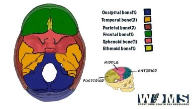
Skull is a bone that protects the brain by forming a protective cavity. It Is formed by many bones joined together by sutures. The number of skull bones is 14. Skull bones are divided into two groups. They are either Cranium and Face. Among the 14 bones, the frontal bone is also one of them.
Cranium is the superior aspect of the skull. Cranium is also called a vault of the l. Cranium is further divided into a Roof and a base. The base of the cranium is further divided into Calvarium and Cranial base.
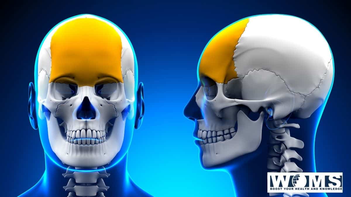
The frontal bone is an unpaired bowl-shaped bone located in the forehead region. It contributes to the formation of the cranium. It lies superior to the nasal bone and anterior to the parietal bone. The frontal bone forms the region of the forehead and resembles a cockleshell in appearance.
Parts of Frontal Bone:
It contains the following parts:
- Vertical part or Squamous part.
- Horizontal parts or Orbital plates, one on each side.
- Nasal part.
Anatomical Position of Frontal Bone:
- Orbital plates look downwards.
- Nasal spine, which is a projection from the nasal part projects downwards and forwards.
Surfaces of Frontal Bone:
- External or Frontal surface.
- Internal or Cerebral surface.
- Right Lateral or Temporal surfaces.
- Left Lateral or Temporal surfaces.
- Right Orbital surfaces.
- Left Orbital surfaces.
1.The external surface:
It is smooth and convex in its general outline. Bounded below by the nasal notch in the middle and the supraorbital margin on each side, above by the parietal border, and on either side by the temporal lines.
Presents:
- Frontal or metopic suture (in 9% of cases): in the lower part in the median plane; indicates the development of the frontal bone in two different segments which fuse into one.
- Frontal tuberosities or eminences: rounded elevations one on each side of the median plane, about 3 cm. above the supraorbital margin; more prominent in young female skulls; form bony landmarks.
- Superciliary arches: arched eminences, one on each side, immediately above the supraorbital margin but separated from the frontal tuberosity by a shallow arched groove. The arch is produced by the frontal air sinus; larger in male than in the female.
- Glabella: a smooth elevation in the median part between superciliary arches.
- Supra-orbital margin: forms on each side upper concave margin of the orbital opening and separate the vertical part from the horizontal part (orbital plate); sharp and prominent in its lateral 2/3 and rounded in its medial 1/3; ends laterally in the zygomatic process which articulates with the frontal process of the zygomatic bone.
Presents:
- Supra-orbital (or foramen or canal) at the junction of lateral 2/3 and medial 1/3. Transmits: Supra-orbital vessels and nerve.
- The foramen for the frontal diploic vein may be present just above or near the supraorbital notch.
- Supra-trochlear or Frontal notch (or foramen): occasionally present medial to the supraorbital notch. Transmits: Supra-trochlear (frontal) vessels and nerve.
Nasal Part: It is a part of the bone projecting downwards below the glabella and between the supra-orbital margins.
Presents:
- Nasal notch: It is an irregular notch that articulates on either side of the midline (from medial to lateral) wit
- nasal bone.
- frontal process of maxilla, and
- lacrimal bone.
- Nasal Spine: a pointed projection downward in the median plane from the lower part of the nasal notch; forms a very small part of the septum of the nose; articulates in front with the nasal crest of the articulated nasal bones, behind with the perpendicular plate of the ethmoid bone and below with the septal cartilage.
- Nasal groove or Nasal surface: lies posteriorly as a very narrow grooved area on either side of the nasal spine; forms a very small part of the roof of the corresponding nasal cavity.
2. The internal surface:
It is deeply concave and lodges the frontal lobe of the brain with its meninges.
Presents:
- Sagittal Sulcus: a vertical groove in the median plane; lodges the anterior part of the superior sagittal sinus; margins of the sulcus give attachment to the falx Cerebri.
- Frontal crest: margins of the sagittal sulcus unite below
to form the crest; it gives attachment to the anterior part of the falx cerebral - Foramen caecum: the frontal crest ends below into a notch which is converted into a foramen by articulation with the alae of the crista Galli of ethmoid bone; usually blind but when a patent, transmits a vein or veins from the nasal mucosa to superior sagittal sinus.
- Granular foveolae: small irregular pits on each side of the sulcus; lodge arachnoid granulations.
- Impressions for cerebral sulci and gyri: on each side of the median plane.
- Furrows for meningeal vessels.
3 & 4Temporal surface:
It is separated from the frontal surface by the superior temporal line; it forms the anterior part of the temporal fossa and presents two temporal lines; the superior line gives attachment to the temporal fascia and the inferior line together with the surface below gives origin to the temporalis muscle.
5 &6 Orbital Surface:
It is the inferior surface of the orbital plate; smooth and concave and forms a major part of the roof of the orbital cavity
Orbital Parts:
Are two thin triangular plates, one on either side of the median plane and forms major parts of the roofs of orbits.
Presents:
- Lacrimal fossa: a shallow depression in the anterolateral part to lodge the lacrimal gland.
- Trochlear fossa or Spine: lies below and behind the medial end of the supra-orbital margin; gives attachment to the fibrocartilaginous pulley of the superior oblique muscle.
Besides the two plates, the orbital part presents:
Presents:
- Ethmoidal notch: a wide U-shaped gap separating the medial margins of the two plates; the medial margins present broken air cells; when the notch is articulated with a cribriform plate of the ethmoid the broken air cells form ethmoidal air sinuses.
- Anterior and Posterior ethmoidal canals: two transverse grooves cross each margin of the notch with similar grooves on the upper surface of the labyrinth of the ethmoid complete the anterior and posterior ethmoidal canals Transmits: Anterior and posterior ethmoidal vessels and nerves respectively.
- The posterior border of each orbital plate: thin and serrated; articulates with the anterior border (of its side) of the lesser wing of the sphenoid bone.
Some terms:
Parietal border (Posterior border) of the vertical part: is thick and strongly serrated; articulates with the frontal borders of the two parietal bones to form the coronal suture. It is continued below into a roughly triangular surface to articulate with the greater wing of the sphenoid bone.
Bregma is the meeting point of two parietal bones and the frontal bone, i.e., the meeting point of coronal and sagittal sutures. It is the site of the anterior fontanelle (unossified membranous part in the fetal skull)
Nasion is the meeting point of two nasal bones with the frontal bone i.e., the meeting point of inter-nasal and frontonasal sutures.
Frontal air sinuses: are two irregular cavities, one on either side of the median plane, within the frontal bone opposite the glabella and superciliary arches; extremely variable on size; roughly triangular; separated from each other by a thin bony septum which may be deviated to one or the other side of the median plane; each communicates with the middle meatus of the corresponding nasal cavity through the frontonasal duct; lined by mucous membrane derived from the nasal cavity
Takeaway :
The frontal bone is one of the skull bones. The cranium of the skull is formed by the frontal bone. Frontal bone has three parts and six surfaces. It also has an important role in protecting and supporting the nervous tissue of the brain. It also helps in giving shape to the skull
Frequently Asked questions
How many frontal bones are there?
The frontal bone is one of the skull bones that resemble a cockleshell. It is one only in number. It consists of three-part i.e is a squamous part, orbital part, and nasal part.
Is frontal bone a pneumatic bone?
The bone that is hollow and consists of many air cells is known as a pneumatic bone. Yes, along with pneumatic bone, it is also a flat bone. Some of the other examples of pneumatic bone are Maxilla, sphenoid and ethmoidal bone.
What’s the importance of frontal tuberosities?
Inferior surfaces of frontal tuberosities correspond with a frontal pole of each cerebral hemisphere.
