The only movable bone in the skull is the mandible. The mandible is the lower jaw of a vertebrate. The mandible can be moved up and down and laterally. This movement is called protraction and retrusion, respectively. Protrusion and retrusion are used synonymously to describe movements of the mandible from an occlusal position towards or away from the midline of the face.
Ossification of the mandible
The only movable bone in the skull, the mandible ossifies during embryonic development. Ossification begins at the endochondral growth plate of the mandibular condyle. As the cartilage calcifies, it forms a bony mass that will become the mandible.
Shapes of the movable bone in the skull
In males, the angle of the mandible is more acute than in females. The angle of the mandible in males is less obtuse than in females. In males, the angle of the ramus is more acute than in females. The height of the corpus is greater in males than in females. In females, the corpus has a longer length than in males. The corpus of males has a wider width than in females. The corpus of males is broader than that of females. The height of the condylar head is greater in males than in females. The height and breadth of the condylar neck are greater in males than in females. The height, breadth, and depth of the glenoid fossae of the temporal bone are greater in males than in females.
Mandible and tooth size
The size of the teeth is related to the size of movable bone in the skull. The alveolar process determines the size of the teeth. The size of the alveolus determines the size of the root of the tooth. The size of the roots of the teeth is proportional to the size of the alveolar process. The size and shape of the teeth are determined by the size and shape of the mandible.
Location of the mandible
The location of the masseter muscle determines the location of the mandible. The masseter muscle is responsible for chewing food. The masseter muscle originates from the mandible and is inserted into the temporal bone. The insertion point of the masseter muscle is located near the middle of the zygomatic arch.
Relations of the mandible
Let’s now have a look at the various relations the only movable bone in the skull.
Muscles
Three muscles control the movement of the mandible. These are the masseter, medial pterygoid, and lateral pterygoid muscles. The masseter muscle acts to protrude the mandible. The medial pterygoid muscle pulls the mandible forward. The lateral pterygoid muscle retracts the mandible. The masseter muscle attaches to the mandible through the mandibular fossa. The medial pterygoid muscle attaches to the mandibular fossa through the infratemporal fossa. The lateral pterygoid muscle attaches to the mandibular ramus through the angular process.
Nerves
The movable bone in the skull contains two main nerves. One nerve runs along the inferior border of the mandible. This is the mental nerve. The other one runs along the superior border of the mandible and divides into four branches. Two branches run on each side of the body of the mandible. They are the buccal nerve and the lingual nerve. The third branch goes towards the lower lip. It is called the mylohyoid nerve. The fourth branch goes towards the tongue. It is called the hypoglossal nerve.
Blood Vessels
Many blood vessels go through the mandible. Many arteries supply blood to the mandible. These include the inferior alveolar artery, the internal maxillary artery, the external carotid artery, the ascending pharyngeal artery, and the facial artery. Many veins drain blood from the mandible. These veins include the external jugular vein, the internal jugular vein, the anterior jugular vein, the posterior auricular vein, the superficial petrosal vein, the transverse sinus, the sigmoid sinus, and the occipital sinus.
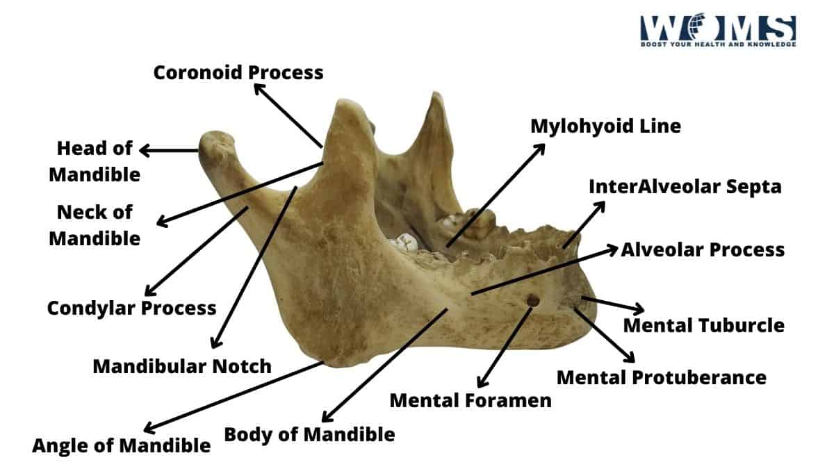
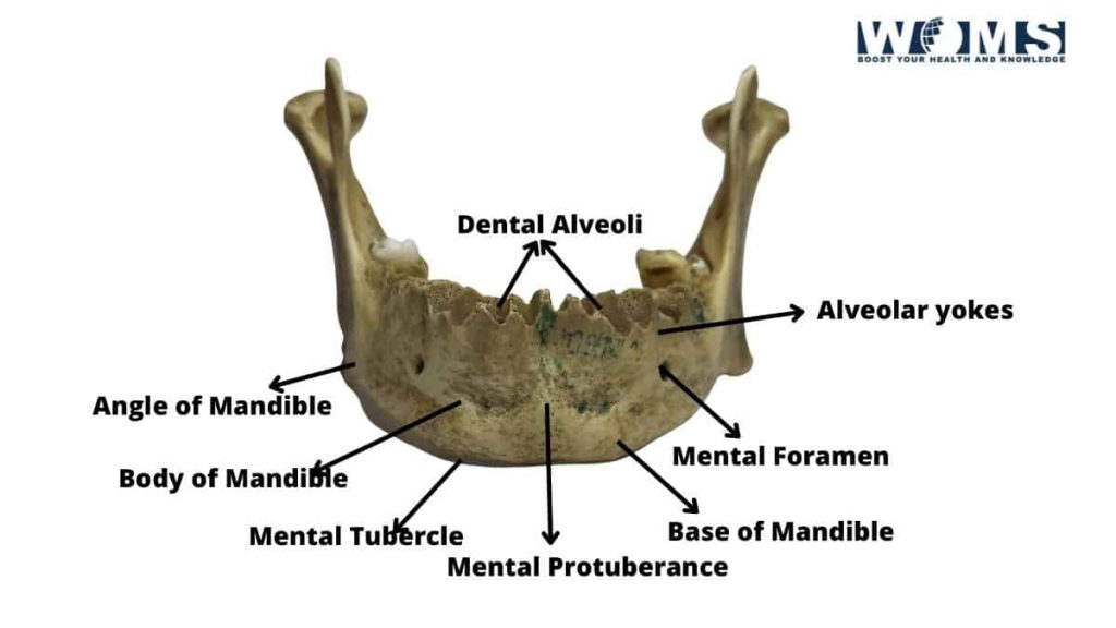
Parts of a mandible
The movable bone in the skull is composed of two parts, the body, and the ramus. The body is in front of the braincase and consists of four parts: the ascending process, coronoid process, angular process, and articular surface. The ramus is a slender bone that connects to the body at its anterior end and articulates with the temporal bone at its posterior end. The mandibular arch is formed by the articular surfaces of the body and ramus. The articular surface of the body forms the inferior border of the mandibular fossa and the superior border of the mental foramen.
Articulation of the mandible
There are five joints in the mandible. These joints are between the following bones: the mandibular condyles and the temporal bone; the mandibular condylar heads and the zygomatic arch; the mandibular symphyses and the hyoid bone; the mandibular rami and the skull base; and the mandibular foramen and the inner ear.
The articular surface on the ramus forms the lateral wall of the oral cavity. The condyle is located on the medial side of the ramus, between the incisura and the glenoid fossa. The movable bone in the skull is attached to the skull via the temporomandibular joint, formed by the mandibular fossae and the glenoid cavities of the temporal bones.
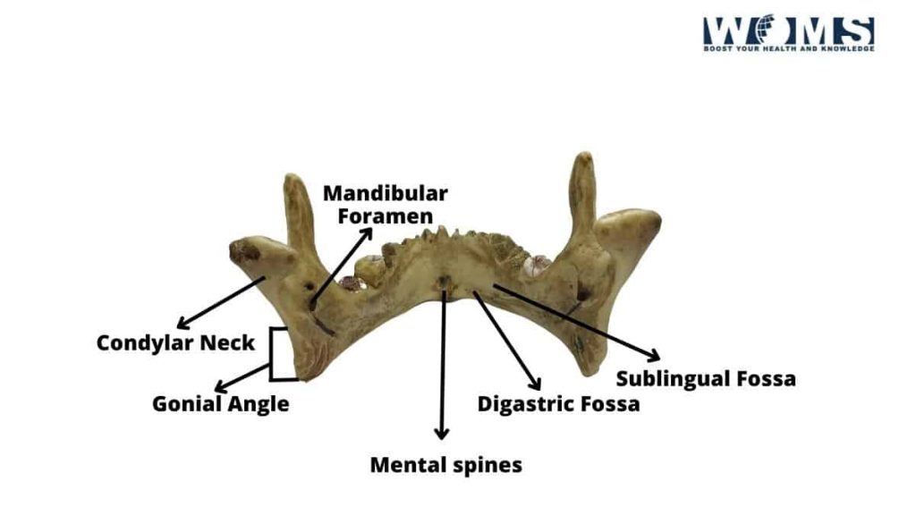
Foramen
The mandible has a large opening called the mandibular foramina. The mandibular foramen is located just below the chin. Through this hole, the trigeminal nerve passes out of the skull. The mandibular canal is another name for this hole. It is a passageway for the spinal cord and the sensory nerves.
Mandibular Canal
The mandibular canal is a long narrow channel running down the length of the mandible. It begins at the mandibular foramens. It ends at the mandibular fossa. The mandibular fossa is where the mandibular nerve leaves the mandible. The mandibular nerve is the largest in the human body. It carries sensation from the face to the brain.
Clinical Relation
The movable bone in the skull is involved in many clinical conditions. Some of these involve the muscles attached to the jaw. Others involve the teeth or their supporting structures. Still, others involve the temporomandibular joints.
Temporomandibular Joint Disorder
A disorder of the temporomandibular joint causes pain and limited jaw motion. This can be caused by trauma, infection, inflammation, tumors, arthritis, congenital deformity, or degenerative disease. Temporomandibular disorders (TMD) affect about 10% of the population. They are more common in women than men. TMDs cause headaches, neck stiffness, fatigue, loss of appetite, soreness in the jaw muscles, toothache, and sensitivity to cold temperatures.
FAQ
What are the different parts of the mandible?
The movable bone in the skull consists of three main parts: the ramus, the corpus, and the coronoid process.
Ramus:
The ramus is the part of the mandible that connects to the rest of the skeleton. It is shaped like an inverted letter U. The inferior border of the ramus forms the posterior margin of the mandible. The superior border forms the anterior margin of the mandible and is connected to the maxilla. The medial surface of the ramus is concave.
Corpus:
The corpus is the part of the jaw that contains most of the tooth sockets. It is shaped like a triangle with rounded corners. The inferior border is convex. The superior border is straight. The left and right sides of the corpus are mirror images of each other.
Coronoid Process:
The coronoid process is located on the medial aspect of the ramus. It is shaped like half of a capital T. Its apex points medially. The coronoid process articulates with the head of the humerus.
How does this movable bone in the skull work?
The mandible works as a lever to help us chew food. When we bite down on something hard, such as a piece of meat, our molars hit the bottom of the jawbone. These molars push up on the top of the jawbone. This pushes the mandible back and up. As the mandible goes up, it rotates around its pivot point at the foramen magnum. This rotation causes the teeth to grind up the food.
What is the function of the mandible in humans?
The mandible helps us eat, speak, hear, smell, and taste. The mandible holds the face upright and allows us to see, breathe, and swallow. It provides support for the tongue and soft palate. The mandible also protects the lower neck and throat from injury.
What is the structure of the mandible in human beings?
The mandible has two bones: the body and the ramus. The body is curved and narrows toward the tip of the chin. The ramus is wider than the body. It extends into the upper part of the temporal bone. The coronoid process projects outwards and downwards. The articular surfaces of the coronoid process articulate with the head of the femur.
Which muscles attach to the mandibular bone?
Many muscles attach to the mandibles. Some of these muscles aid in chewing. Others are involved in opening and closing the mouth.
Describe the movements of the mandible.
The mandible moves when you open your mouth or close it. You can move the mandible by moving your tongue, lips, cheeks, and jaw.
Conclusion
The movable bone in the skull is one of the important bones in the human body. It comprises many pieces that form a complex system of levers. The mandible acts as a lever when you bite down on something hard. When you do so, your molars hit the bottom edge of the jawbone. Your molars then push up on the top edge of the jawbone, which moves the entire mandible backward and upwards. This movement is what makes the jaw move back and forth.