Parietal Bone
Parietal Bone: One Of the skull bone
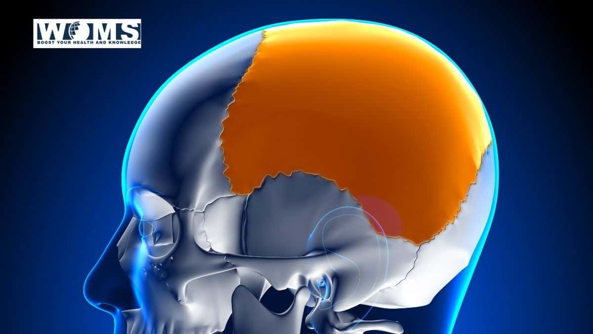
Skull is part of the skeleton. It supports the part of the face and forms a cavity for the brain. Skull is formed by many bones joined together by sutures. Skullbone is made up of 22 bones. There are 8 cranial bones and 14 facial bones that form the skull. Out of 8 cranial bones of the skull bone, 2 of them are Parietal bones. Parietal bones are bilateral bones. It forms the side and roof of the cranium. There is also the presence of Parietal eminence and Parietal foramen.
The parietal bone is two in number, situated between the frontal bone in front and occipital bone behind. It is irregularly quadrilateral in shape. This bone overlies the parietal bone of the brain. The two together form the greater part of the vault and the sides of the skull.
Parts of Parietal Bone
1.Surfaces :
- (i) External
- (ii) Internal
2. Borders :
- (i) Superior (sagittal),
- (ii) Inferior (squamosal).
- (iii) Anterior (frontal), and
- (iv) Posterior (occipital).
3. Angles:
(i) Antero-superior (frontal),
(ii) Antero-inferior (sphenoidal),
(iii) Postero-superior (occipital) and
(iv) Postero-inferior (mastoid).
Determination of side :
- The superior (sagittal) border which is straight, thickly serrated
and the longest lie above in the median plane. - The external surface is convex.
- The inferior border is arched.
- Antero-Inferior (sphenoidal) angle is acute.
Surfaces of Parietal Bone
External surface :
The external surface of the parietal bone is smooth and convex in all directions.
Presents:
(i) Parietal tuberosity (eminence):
A parietal eminence is a rounded eminence near the center. The point of ossification is denoted by parietal eminence
Importance :
- forms an important bony landmark.
- corresponds to the maximum transverse diameter of the skull.
- corresponds to the upper end of the posterior ramus of the lateral sulcus of the cerebral hemisphere.
opposite this lies the center for written and printed words. - The Centre of ossification first starts at this
point.
(ii) Temporal lines :
Two arched lines, one above the other,
known as the superior and inferior temporal lines, cross this surface from before backward.
(a) Superior temporal line: traverses the middle of this surface and gives attachment to the temporal fascia. The area above this line is covered by the galea aponeurotica (epicranial aponeurosis).
(b) The inferior temporal line and the area below it give origin to the temporalis muscle; the area below this line forms part of the temporal fossa.
iii) Parietal foramen:
The parietal foramen lies near the posterior part of the superior border. The parietal emissary vein opens through the parietal foramen. It is always not present.
Transmits :
(a) a small branch of the occipital artery.
(b) an emissary vein connecting the superior sagittal sinus with the occipital vein.
- Internal (Cerebral) surface: The internal surface of the parietal bone is deeply concave.
Presents:
(i) Sagittal sulcus: a longitudinal shallow groove along the superior (sagittal) border which with a similar groove of the opposite bone forms the sagittal sulcus: lodges the superior sagittal sinus; margins of the sulcus give attachment to the falx Cerebri
(ii) Granular foveolae: small irregular pits on each side of
the sulcus: lodge arachnoid granulations.
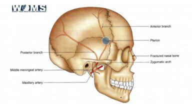
(iii Groves for branches of middle meningeal vessels: the impression for the anterior branch begins at or near the anteroinferior angle and that of the posterior division at a close distance from the posteroinferior angle.
(iv) Impressions for cerebral gyri.
Borders of Parietal Bone
1. Superior (sagittal) border: straight, thickly serrated, and the longest border; articulates with the corresponding border of the opposite parietal bone to form the sagittal suture
2. Inferior (squamosal) border: arched and divided into 3 parts.
- Anterior: beveled at the expense of its outer surface and articulates with the greater wing of the sphenoid.
- Intermediate: arched and beveled at the expense of its
the outer surface and articulates with the squamous part of the temporal bone. - Posterior: straight and thickly serrated; articulates with
the mastoid part of the temporal bone.
- Anterior (frontal) border: thick and deeply serrated; beveled at the expense of its inner surface above and the outer surface below, articulates with the posterior (parietal) border of frontal bone to form one half of the coronal suture.
- posterior (occipital) border: also thick and serrated; articulates with the parietal (lambdoid) border of the squamous part occipital bone to form one half of the lambdoid suture.
Lambda: is the meeting point of two parietal bones and occipital bone, i.e., the meeting point of sagittal and lambdoid sutures. It is the site of the posterior fontanelle (unossified membranous part) in the fetal skull.
Angles:
- Antero-superior (frontal) angle: formed by the meeting of sagittal and frontal borders: corresponds with bregma (meeting point of sagittal and coronal sutures).2. Antero-inferior (sphenoidal) angle: thin and most pointed. It is formed by the meeting of frontal and squamosal borders: corresponds with pterion. The pterion is the meeting place of 4 bones-frontal. parietal,
sphenoid (its greater wing), and temporal (it’s a squamous part), at the sphenoidal angle of the parietal bone. It is an important bony landmark.
Importance:
- Opposite this point lies:
- the anterior division of the middle meningeal artery.
- the division of the stem of the lateral sulcus of the cerebral hemisphere into its three rami.
- Broca’s motor speech center of the brain.
- Postero-superior (occipital) angle: formed by the meeting of sagittal and occipital borders, corresponds with lambda.
- Postero-inferior (mastoid) angle: blunt; formed by the meeting of occipital and squamosal borders; corresponds with asterion.
Asterion: is the meeting place of 3 bones-parietal, occipital, and temporal (it’s a mastoid part) at the mastoid angle of parietal bone; on its internal surface lies a sulcus where the transverse sinus ends and the sigmoid sinus begins.
Takeaway:
The parietal bone is two in number, situated between the frontal bone in front and occipital bone behind. It is irregularly quadrilateral in shape. This bone overlies the parietal bone of the brain. The two together form the greater part of the vault and the sides of the skull. It articulates with the other five bones like the frontal, sphenoid, temporal, occipital, and contralateral parietal bones. The Parietal bone protects the underlying brain structure i.e parietal lobe.
If you would like to know more about other skull bones then do read: Frontal bone