The complete guide to vertebral artery anatomy
The vertebral artery is the major blood vessel of the spine. It supplies blood to the spinal cord and runs along the vertebrae. It passes through the thoracic cavity and enters the lumbar region.
The vertebral artery has many branches, and it also has several valves, which help prevent it from becoming blocked or impeded.
Table of Contents
Location of the vertebral artery
The vertebral artery is found in the upper part of the neck. It runs through the spine and branches out into many different arteries. Since it is a large blood vessel, many valves help to prevent it from becoming blocked or impeded.
A blocked or occluded blood vessel is a serious cause of serious damage to the body. The reason behind this problem is atherosclerosis, which can develop due to low-density lipoprotein (LDL) cholesterol and high-density lipoprotein (HDL) cholesterol levels in the blood.
The extent of the vertebral artery
The vertebral artery terminates between T1-2 in front of the medulla oblongata, where it bifurcates into two branches that run to either side of the brainstem. These arteries then subdivide into branches supplying blood to different regions; these smaller arteries are called anterior spinal, posterior spinal, and craniocervical arterial trunks.
What are the different systems of the vertebral artery?
The different systems of the vertebral artery can be broken down into the following three categories:
- The first system is called the anterior aortic arch. It includes the left and right common carotid arteries, internal carotid arteries, and vertebral arteries.
- The second system is called the posterior aortic arch. It includes the left and right subclavian arteries and vertebral arteries.
- The third system is called the vertebral body artery system. It includes the left and right posterior cerebral arteries, vertebral artery, posterior communicating artery, and basilar artery.
What are some important landmarks on the vertebral artery?
The vertebral artery is a major artery that runs along the spine. It begins near the base of the skull and extends down to the tailbone, branching into two large arteries just before it passes through each side of the spinal cord.
It is a large artery and a main central supply to the brain and spinal cord, but it passes through very small openings into the skull. No one knows where this artery originates, but some researchers think it may begin in the first ventricle of the brainstem or perhaps in the floor of the third ventricle.
Various methods have been used to study vertebral arteries because it is difficult to locate them accurately in these small spaces, and it is difficult to perform dissections on them.
Symptoms of a damaged or compromised vertebral artery
A damaged or compromised vertebral artery symptoms are visible through myelopathy and include progressive motor weakness in the lower extremities. In some cases, there may be other neurological signs such as an absent reflex in the lower extremities, pain and tingling, and leg weakness in the spinal cord region.
Is it possible to identify a compromised or damaged vertebral artery through imaging tests?
The vertebral artery is a structure that can be identified on MRI imaging using a contrast agent. Also, there are other ways to diagnose this type of disease: angiography (to examine the blood vessels), magnetic resonance angiography (MRA) (to examine the flow of blood through blood vessels), CT, CAT scan, and nuclear medicine scans such as single-photon emission computed tomography (SPECT) and positron emission tomography (PET).
What is the function of the vertebral artery?
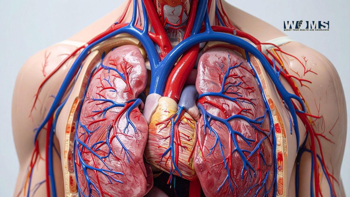
The vertebral artery carries blood to the brain and spinal cord base, which are vital structures that make up much of the central nervous system. It is one of the two terminal branches of the subclavian artery and arises opposite the cervical vertebrae at C6-7. The subclavian arteries are located under collarbones and give birth to different arteries throughout their journey toward their terminus at C1-2, where they bifurcate into left and right common carotid arteries.
Which portion of our spine does it service?
The spinal cord. It’s the lifeline of our nervous system, supplying messages to and from various parts of our body. The vertebrae are the bones that protect this delicate nerve bundle, which is about three and a half inches long and located in the middle of your back. It sits behind your neck, at the base of the skull.
How does it provide blood flow to the brain?
The vertebral artery is a large artery that supplies blood to the head, neck, and upper trunk. It is one of the two terminal branches of the subclavian artery and arises opposite the cervical vertebrae at C6-7. The subclavian arteries are located under collarbones and give birth to different arteries throughout their journey toward their terminus at C1-2, where they bifurcate into left and right common carotid arteries.
What are some other names for the vertebral artery?
The vertebral artery is also known as the basilar artery, The vertebral artery is located in the posterior cervical region of the neck and is formed by a bifurcation of the common carotid artery. It is named because it gives rise to several other branches, which travel along the vertebral column. The arteries that travel along the vertebral column are the anterior spinal, posterior spinal, and craniocervical arterial trunks.
How can I reduce the effect of a blocked or damaged Vertebral Artery?
A blockage or damage can be reduced by taking medication such as aspirin while avoiding many things that cause stress and emotion, such as smoking and heavy exercise. However, other medications may not be recommended if there are signs of blood clotting or bleeding or someone has a heart attack. If a blockage or damage occurs, it is best to consult a physician to help with treatment.
What is Vertebrobasilar Insufficiency?
Vertebrobasilar Insufficiency is a condition in which the vertebral arteries and basilar artery are narrowed or blocked, leading to impaired blood flow to the brain. This condition can be caused by multiple factors such as:
How is it diagnosed?
It is usually diagnosed through angiography. To do so, a contrast dye will be injected into the bloodstream, which can then be used to see any blockages in the artery(s). A CT scan or an MRI will show if the arteries are completely blocked. This test is also used to see how long it takes for blood to return after a blockage.
What Is Vertebral Artery Bypass Surgery? How Is It Involved & How Does It Work?
This is a very important surgery, which is very rarely done. It involves the removal of a large section of the vertebrae to bypass an artery that supplies blood to the brain. This surgery can be done on both men and women, and it is usually combined with other surgeries to help treat certain other conditions.
The surgeon removes part of the vertebrae by cutting through them and taking out a piece of bone at each end. He then inserts an external access tube into this area, which lets him make sure that the bone from one side does not get in the way when he makes his incision on the other side. The surgeon then takes out a section of bone from each end, which acts as an anchor for all these pieces of bone.
Vertebral artery bypass surgery is a type of surgery used to treat people with severe heart disease.
How is vertebral artery bypass done?
It is carried out by inserting a new artery from the lower back into the upper part of the spine, where it will bypass the diseased area and supply blood to the heart. This procedure is safe and effective, but it can be difficult to perform as it requires extensive spinal surgery.
A procedure that can be performed safely and economically is a spinal surgery called L1-L5 fusion. This surgery is used to correct the abnormal curves in the spine. L1-L5 fusion avoids the risk of sciatica, pinched nerves, or herniated discs by correcting these abnormalities with only one operation.
L1-L5 fusion is a technique orthopedic surgeons use to reduce the risk of herniated discs. The procedure is used to correct the abnormal position of one or more spinal segments during surgery. In this case, the surgeon will perform a fusion of the L1 vertebra and the L2 vertebra. This allows for proper alignment of all levels in the spine by correcting an abnormal
Vertebral artery and his Relation to Artery Surgery
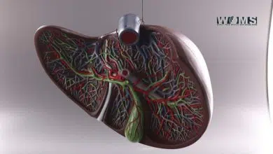
The vertebral artery is a major blood vessel in the spine. It carries blood from the heart to the brain. The vertebral artery is created by a small branch of the internal iliac artery, and it supplies blood to the spinal cord and brain.
Vertebral artery bypass surgery uses surgeons to create an artificial artery by removing part of an existing vertebral artery. This procedure is used in cases where there are tumors or other problems in that area of the body that could cause problems with blood flow to that area.
How can I Avoid Dying from Bleeding Disorder?
A bleeding disorder is a medical condition in which the blood vessels in the body leak and become inflamed. It can be caused by various diseases such as anemia, leukemia, or thrombosis.
A typical scenario in a hospital. Bleeding disorders are caused when blood clots form in the vessel walls of your body. A patient is being treated with chemotherapy, and the treatment is not going well. The patient dies within a few weeks.
Bleeding disorders are serious medical conditions that can lead to death. If you have bleeding disorders, you must take the necessary steps to treat them.
How Vertebral Artery Bypass Surgery Works
This section is about a procedure that involves the vertebra and the bypass. The vertebra is an important structure in the spine. It connects the two bones of your back. The bypass is a small tube that helps move blood through your body.
The bypass is a small tube that provides the blood with the necessary oxygen and nutrients to prevent or relieve many diseases.
The bypass is a small tube that provides the blood with the necessary oxygen and nutrients to prevent or relieve many diseases. It is a lifesaver for those suffering from high blood pressure, hypertension, and other chronic conditions requiring frequent hospitalizations.
How the High-Risk Procedure Can Save Your Life in the Event of an Incapacitation or Injury
Your spinal cord is the part of your body that controls your movement, posture, and other functions. It is not only important for your physical health but also for the quality of life you enjoy.
The vertebra procedure is a surgical procedure that treats spinal cord injuries or spinal cord vulnerability. The procedure involves removing the damaged vertebrae and replacing them with ones from another part of the spine to avoid further damage to the spinal cord.
This procedure involves removing the damaged vertebrae and replacing them with ones from another part of the spine to avoid further damage to the spinal cord. This procedure is considered a risky operation and requires surgical expertise, but the benefits of this surgery outweigh its risks. The surgeon will remove cancerous cells from a patient’s spine. He will then replace them with healthy ones.
Conclusion
It is one of the 24 primary arteries in the human body. Its blood supply comes from the inferior cervical spine and the cranial part of the internal carotid artery, which carries blood from the brain and nose, respectively. The left vertebral artery descends from the internal carotid artery in the brainstem through a short passage called the pons, where it divides into two branches at each level of that region. Each branch then carries its component to one side of the brain.
FAQs
Where is the vertebral artery located?
The vertebral artery is located on the posterior side of the spinal column and runs from the skull base to C1. It is a continuation of the subclavian artery.