Veins of upper limb
Introduction of the upper limb with veins of upper limb
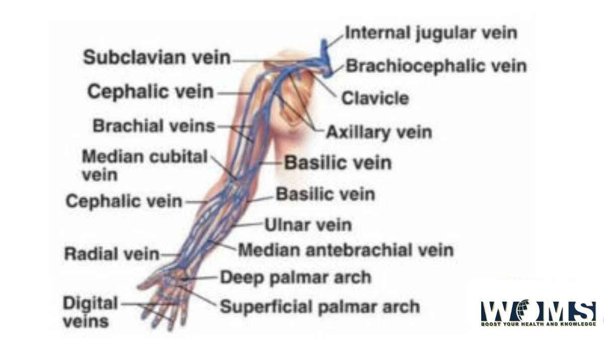
The region of our body, which extends from the deltoid region and includes hands, arms, shoulder, and axilla is called the Upper limb. The upper limb consists of Hands, Arms, and Forearm. Veins of the upper limb are essential to let’s discuss it. Veins of arms are Cephalic vein, Basilic vein, and Median Cubital vein.
As we all know, the human body comprises bone, muscle, nerves, arteries, veins, and many more. So we are going to discuss detail about the veins of the upper limb. Veins of the upper limb consist of arm veins, and forearms veins. This article will give you all the necessary information about the veins of the arm.
There are two sets of veins in the upper extremity. These include superficial as well as deep veins. The superficial veins are present between two layers of superficial fascia just beneath the integument. On the other hand, the arteries accompany the deep veins. Valves are present on both superficial and deep veins. However, the number of valves in deep veins is more as compared to the superficial veins.
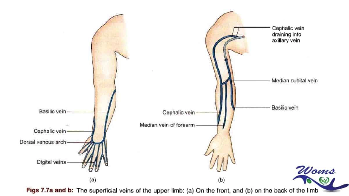
Table summary of the veins of the upper limb
| Hand | Superficial palmar arch
Deep palmar arch |
| Arm | Cephalic vein
Basilic vein Median forearm vein |
| forearm | Brachial veins
Basilicaveins Cephalic vein |
| shoulder | Axillary vein
Subclavian vein |
Superficial veins
Superficial veins of the upper limb assume importance in medical practice because these are most commonly used for intravenous injections and for withdrawing blood for testing.
General Remarks
- Most of the superficial veins of the limb combine two large veins, cephalic (preaxial) and basilic (postaxial). An accessory cephalic vein is often present.
- The superficial veins run away from pressure points. Therefore, they are absent in the palm (fist area), along the ulnar border of the forearm (supporting border), and in the back of the arm and trapezius region (resting surface). This makes the course of the veins spiral, from the dorsal to the ventral surface of the limb.
- The preaxial vein is longer than the postaxial. In other words, the preaxial vein drains into the deep (axillary) vein more proximally (at the root of the limb) than the postaxial vein, which becomes deep in the middle of the arm.
- If a vein becomes deep in early, then it is better because the muscular compression aids the venous return. The load of the preaxial (cephalic) vein is greatly relieved by the more efficient postaxial (basilic) vein through a short-circuiting channel (the median cubital vein situated in front of the elbow) and partly also by the deep veins through a perforator vein connecting the median cubital with the deep vein.
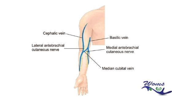
- Cutaneous nerves and superficial lymphatics accompany the superficial veins. Arteries don’t accompany the superficial veins. The superficial lymph nodes lie along the veins and the deep lymph nodes along the arteries.
- For intravenous injections, superficial veins are mostly used.
Cephalic Vein
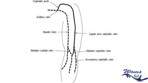
One of the superficial veins of the upper limb is the cephalic vein. The cephalic vein is the preaxial vein of the upper limb (cf. great saphenous vein of the lower limb). It begins from the lateral end of the dorsal venous arch.
It runs upwards:
- Through the roof of the anatomical snuff box,
- Winds around the lateral border of the distal part of the forearm
- Continues upwards in front of the elbow and along the lateral border of the biceps brachii,
- Pierces the deep fascia at the lower border of the pectoralis major,
- Runs in the deltopectoral groove up to the infraclavicular fossa, where
- It pierces the Clavipectoral fascia and joins the axillary vein.
At the elbow, the more significant part of its blood is drained into the basilic vein through the median cubital vein, and partly also into the deep veins through the perforator vein.
It is accompanied by the lateral cutaneous nerve of the forearm and the terminal part of the radial nerve. An accessory cephalic vein is sometimes present’ It ends by joining the cephalic vein near the elbow.
Basilic Vein
The basilic vein is the postaxial vein of the upper limb (cf. short saphenous vein of the lower limb).
Basilica vein begins from the medial end of the dorsal venous arch.
It runs upwards:
- Along the back of the medial border of the forearm,
- Winds around this border near the elbow,
- Continues upwards in front of the elbow (medial epicondyle) and along the medial margin of the biceps brachii up to the middle of the arm, where
- It pierces the deep fascia, and
- Runs along the medial side of the brachial artery up to the lower border of the teres major, where it becomes the axillary vein.
About 2.5 cm above the medial epicondyle of the humerus, it is joined by the median cubital vein. The basilic vein is accompanied by the posterior branch of the medial cutaneous nerve of the forearm and the terminal part of the dorsal subdivision of the ulnar nerve.
Median Cubital Vein
The median cubital vein is a sizeable communicating vein that shunts blood from the cephalic to the basilic vein. The Median Cubital vein begins from the cephalic vein 2.5 cm below the bend of the elbow, runs obliquely upward and medially, and ends in the basilic vein 2.5 cm above the medial epicondyle.
The brachial artery is separated from it by the bicipital aponeurosis.
The median cubital vein may receive tributaries from the front of the forearm (median vein of the forearm). The median cubital vein is connected to the deep veins through a perforator vein, which pierces the bicipital aponeurosis.
The perforator vein fixes the median cubital vein and thus makes it ideal for intravenous injections.
Digital veins ( Veins of the digits )
The dorsal digital veins pass along the sides of the fingers and are connected by communicating oblique branches. Veins that are present on the adjacent side of the fingers join together. Thus there is the formation of three dorsal metacarpal veins, which end in a dorsal venous network opposite the center of the metacarpal. The radial part of the network is joined by the digital dorsal vein from the radial side of the index finger and the digital dorsal veins of the thumb and extends upward like the cephalic vein.
The ulnar part of the network receives the dorsal digital vein from the ulnar side of the little finger and continues upward like the basilic vein. A communicating branch often connects the dorsal venous network to the cephalic vein around the center of the forearm.
Then there are volar digital veins in each finger that remain connected to the dorsal digital veins by inter-capitular oblique veins. They drain into a venous plexus located above the lateral and hypothenar eminences and through the front of the wrist.
Median vein of the forearm
The median vein of the forearm begins from the palmar venous network and ends in any one of the veins in front of the elbow, mostly in the median cubital vein.
Deep Veins
Deep veins start as small venae comitantes running on each side of digital veins. These continue proximally as superficial and deep palmar arches. The deep arm veins will be discussed further.
Then, these courses proximally continue as venae comitantes of radial and ulnar arteries; which further join to form the brachial veins. Brachial veins lie on each side of the brachial artery.
These join the axillary vein at the lower border of the teres major. The axillary vein is described in the axilla.
The deep veins of the upper limb are discussed below:
The brachial veins are placed on each side of the brachial artery, receiving tributaries corresponding to the detached branches from that vessel. Just close to the lower margin of the subscapularis, they join the axillary vein wherein the medial often joins the basilica vein. There are numerous anastomoses of the deep veins with each other as well as with the superficial veins. Next in this article of veins of the arm, including all the veins of the upper extremity is an axillary vein.
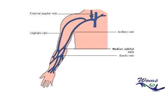
The axillary vein begins on the lower edge of the Teres major, as the continuation of the basilica vein increases in size as it rises and ends at the outer edge of the first rib as a subclavian vein. Near the lower edge of the subscapularis, it receives the brachial veins and, near its termination, the cephalic vein. Its other tributaries correspond to the branches of the axillary artery.
It is located on the medial side of the artery, which partially overlaps between the two vessels are the medial cord of the brachial plexus, the median, the ulnar, and the medial anterior thoracic nerves. It is equipped with a pair of valves opposite the lower edge of the subscapularis; The valves are also located at the ends of the cephalic and subscapular veins.
The subclavian vein, the continuation of the axillary axis, extends from the outer edge of the first rib to the sternal end of the clavicle, where it joins the internal jugular to form the unnamed vein. It is related, opposite, to the clavicle and the Subclavian, behind and above, with the subclavian artery, from which it is separated medially from the anterior scalene and the phrenic nerve. It then rests in a depression in the first rib and pleura. It usually comes with a pair of valves, which are approximately 2.5 cm apart.
Clinical anatomy of the veins of the upper limb
The median cubital vein is the vein of choice for intravenous injections, for withdrawing blood from donors, and for cardiac catheterization, because it is fixed by the perforator and does not slip away during piercing. When the median cubital vein is absent, the basilic is preferred over the cephalic because the former is a more efficient channel. The basilic vein runs along a straight path, whereas the cephalic vein bends acutely to drain into the axillary vein.
The cephalic vein frequently communicates with the external jugular vein utilizing a small vein crossing in front of the clavicle.
In operations for removal of the breast (in carcinoma), the axillary lymph nodes are removed, and it sometimes becomes necessary to remove a segment of the axillary vein.
These cases/ the communication between the cephalic vein and the external jugular vein enlarges considerably and helps in draining blood from the upper limb.
In the case of fracture of the clavicle, the communication channel rupture may lead to the formation of a large hematoma, i.e., collecting blood.
The above article is a description of the veins of the upper limb. All the necessary information on the major veins of the upper limbs with a detailed explanation of the veins of the arm is included in this article. All the arm veins were also discussed simply. We hope that this article will help you learn about the veins of the upper limb properly.
Takeaway:
Veins of the upper limb consist of arm veins, and forearms veins. There are two sets of veins in the upper extremity. These include superficial as well as deep veins. The superficial veins are present between two layers of superficial fascia just beneath the integument. On the other hand, the arteries accompany the deep veins. Valves are present on both superficial and deep veins. However, the number of valves in deep veins is more as compared to the superficial veins.
Do read: Phlebitis and Thrombophlebitis
FAQs:
Which veins are present in the upper limb?
The superficial veins like cephalic and basilica veins are present in the upper limb.
What are the main veins of the upper limb from which blood is drawn out?
Blood is drawn out from the three veins. These three veins are the median cubital vein, cephalic, and basilica vein.
Which is the most common vein used to draw out the blood?
The blood is drawn out commonly from the median cubital vein.
Which is the largest vein present in the arm?
The antecubital vein also called as median cubital veins is the largest vein present in the arm.