Tongue Muscles Anatomy and Important Clinical Considerations
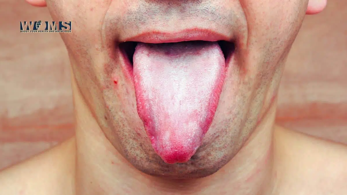
The tongue represents itself as a form of smooth muscle. Most people wonder about the various functions performed by this chunk of smooth muscle. There are different tongues muscles that help to perform particular motor movements. Besides being a motor system due to muscles, the tongue also offers several sensory functions like the taste, touch, etc. Let us look at the anatomical as well as physiological features of tongue muscles to know in detail.
Tongue muscles are divided into two major groups. These are known as:
- Intrinsic muscles
- Extrinsic muscles
Intrinsic tongue muscles:
Intrinsic tongue muscles are those that are located within the tongue with no attachments outer of the tongue. These muscles have origin and insertion points inside the tongue. There are four pairs of intrinsic tongue muscles that fall in this category. These are named according to the direction they come forward.
- Superior longitudinal lingual muscle – This tongue muscle helps to reduce the overall length of the tongue. Moreover, it helps to curl the tongue in an upward direction.
Origin – it originates from the median fibrous septum closer to the epiglottis.
Insertion – it inserts near the anterolateral edges of the tongue.
- Inferior longitudinal lingual muscle – This tongue muscle also helps to reduce the length of the tongue by curling the tongue in a downward direction.
Origin – it originates from the root of the tongue.
Insertion – it inserts into the tip point or apex of the tongue.
- Transverse lingual muscle – This tongue muscle works opposite previous longitudinal muscles. It helps to increase the length of the tongue, making it narrower than normal.
Origin – it originates from the same point where superior longitudinal lingual muscle, median fibrous septum.
Insertion – it inserts along the lateral sides of the tongue.
- Vertical lingual muscle – This tongue muscle works to flatten the tongue, making it wider.
Origin – it originates from the submucosal fibrous layer, located at the bottom of the tongue.
Insertion – This tongue muscle inserts into the edges of the tongue present in the bottom.
Function:
Though, every intrinsic tongue muscle performs a separate function. But, overall, these muscles are responsible for:
- Maintaining the shape (narrow or wider) and size of the tongue
- Play the main role while eating, swallowing, and talking
How do these tongue muscles perform these motor movements?
Intrinsic muscles have a specific motor innervation to perform these tongue movements like tongue rolling, etc. Cranial nerve XII, the hypoglossal nerve, innervates these intrinsic tongue muscles to perform these motor functions.
Extrinsic tongue muscles:
Extrinsic tongue muscles are those that usually originate from the outer surface of the tongue. But, these muscles show direct attachment or insertion point to different other structures of the mouth. There are also four pairs of extrinsic tongue muscles that perform several functions.
Function:
These extrinsic tongue muscles also perform multiple tongue movements but are different from the intrinsic tongue muscles. They perform the following tongue movements.
- Protrusion
- Retraction
- Elevation
- Depression
Let us look at these extrinsic tongue muscles to know in detail.
- Genioglossus – this tongue muscle seems like a fan-shaped muscle. It originates from the superior mental spine located above the posterior aspect of the mandible. At this point, the inferior fibers insert into the hyoid bone. Whereas, the other fibers move upward along the overall length of the dorsum of the tongue. Here, these fibers terminate with blending in the intrinsic muscle fiber.
Origin – superior mental spine on the posterior surface of the mandible
Insertion – moves along the dorsum of the tongue and inserts into the body of the hyoid bone.
Function:
- Bilateral contraction of genioglossus muscle helps in the depression of the tongue, protruding the tongue outside the mouth (inferior and middle fibers).
- Unilateral contraction helps to turn the tongue contralaterally
- Hyoglossus – this tongue muscle looks like a quadrangular muscle, originating from the greater horn and body of the hyoid bone. It extends in a forward direction through the oropharyngeal triangle and inserts into the ventral surface of the lateral tongue.
Origin – it originates from the greater horn and body of the hyoid bone.
Insertion – it inserts into the inferior surface of the lateral tongue.
Function:
- It is a broader tongue muscle forming the floor of the oral cavity.
- It depresses the lateral surface of the tongue with retrusion of the tongue.
- Styloglossus – This tongue muscle is in the form of a thin strip originating from the anterolateral surface of the styloid process of the temporal bone. Longitudinal part of this muscle blends with the fibers of inferior longitudinal muscles. Whereas, superior fibers blend with the fibers of hyoglossus.
Origin – it originates from the styloid process of the temporal bone and stylomandibular ligament
Insertion – inferior fibers terminate as blending in inferior longitudinal muscle
Superior fibers insert into the fibers of the hyoglossus muscle
Function:
- This tongue muscle contracts with performing the retraction or retrusion of the tongue.
- In addition, it also helps to elevate the lateral aspects of the tongue
- Palatoglossus – This tongue muscleappears as a bow-shaped muscle connected with both the tongue and soft palate. This tongue muscle originates from the palatine aponeurosis of the soft palate with downward extension laterally. Ultimately, it gets inserted into the dorsal surface of the tongue blending within intrinsic muscles.
Origin – it originatesfrom the palatine aponeurosis or union point of the soft palate.
Insertion – it inserts into the lateral surface of the tongue
Function:
- This tongue muscle helps to coordinate the movements of the soft palate and tongue.
- In addition, it helps in the elevation of the root of the tongue and constriction of the isthmus of fauces.
You should check this detailed article on Pharynx anatomy to know more about this.
Innervation of Extrinsic tongue muscles:
These extrinsic tongue muscles perform various tongue movements with the help of nerve supply. All of these extrinsic muscles are supplied by cranial nerves. Genioglossus, hyoglossus, and styloglossus receive motor innervation by the cranial nerve XII, the hypoglossal nerve.
Whereas, Palatoglossus receives its motor innervation through the cranial nerve X, vagus nerve, via pharyngeal plexus.
Blood supply to the tongue muscles:
The lingual artery, a branch of the external carotid artery, offers the blood supply to most of the tongue structure. In addition, the tonsillar artery, a division of the facial artery, also provides collateral blood supply to the tongue musculature.
Lymphatic drainage:
There are different lymph nodes present that drain the lymph from the tongue.
Anterior two-thirds – drains into the submental and submandibular lymph nodes, which drain into the deep cervical lymph nodes
Posterior third – drains directly into the deep cervical lymph nodes
Clinical considerations of the tongue:
Tongue offers multiple clinically important points that are helpful in diagnosing several clinical problems. Let us look at these medically important points to appreciate the use of information in clinical practice.
Tongue-tie:
It is a medical condition in which the anteroinferior surface of the tongue remains connected to the midline frenum through connective tissue. Usually, apoptosis occurs at an early age to resorb this connection. But, If it fails, this condition presents as tongue-tied in children. It may cause problems in phonetics and breastfeeding. Usually, a surgical procedure, frenuloplasty, is performed to reposition the attachment.
Damage to tongue muscles:
Some traumatic injuries may occur that can damage the tongue muscles to a different extent. Damage to the tongue muscles may affect speech and masticatory function. Penetrating trauma to the tongue muscle may lead to the temporary failure of tongue movements. As the tongue tissue heals, tongue movements start becoming normal.
Damage to the Hypoglossal nerve:
All tongue muscles except palatoglossus are innervated by the hypoglossal nerve. Any damage to the hypoglossal nerve may lead to the loss of motor function of these muscles. The hypoglossal nerve does not exhibit decussation. Therefore, the damage appears on the same side on which the nerve injury occurred. Unilateral damage to the nerve may result in the muscular reduction, occurring on the same side of the tongue. Unilateral injuries usually don’t affect speech and mastication.
Bilateral injuries present as the generalized paralysis of the tongue, known as dysarthria. Moreover, the tongue lost its function as mastication.
We also suggest everyone to clean your tongue regularly. You should check this detailed article on Tongue cleaner types and uses to know more.
Frequently asked questions (FAQs)
How can we check the weakness of the tongue?
It is usually checked clinically through the help of a tongue depressor. Doctors usually ask the patient to push the tongue against a tongue depressor held vertically at a slight distance from the patient’s lips.
What is tongue paralysis?
Tongue paralysis usually occurs due to an injury or trauma to the hypoglossal nerve fibers. In this condition, patients fail to perform different tongue movements.