Diagram of Muscle Fiber: 3 Types, Functions, and Anatomy
A Diagram of muscle fiber is an effective way to understand the muscles of the human body. The whole body’s musculature performs various functions in daily life. In addition, they help orient the skeletal movements, physical contraction, and blood pumping outside the heart. Moreover, every body’s structure is associated with a different type of muscle. Muscles contain a single structural unit known as muscle fiber. This article clears the basic concepts related to the diagram of muscle fiber. Moreover, The diagram of muscle fiber orients the details about physiology, types, and different other aspects.
Muscle contains multiple and different types of muscle fibers, cylindrical in shape and ranging from a diameter of 0.02 to 0.08mm as seen in the diagram of muscle fiber. Muscle fibers may run along the whole length of the muscle. Its size may vary up to several tens of centimeters. Whereas in contrast, several muscles contain tendons at every edge. And these muscle fibers move diagonally between the tendons but across the muscle. This can be seen in a diagram of muscle fiber.
There may be multiple variations in the orientation of muscle fibers depending upon the function of the muscle. There are several types of muscle fibers exhibiting different kinds of features. Let us have a look at the detailed anatomy of the diagram of muscle fiber to learn.
What are the different types of muscles?
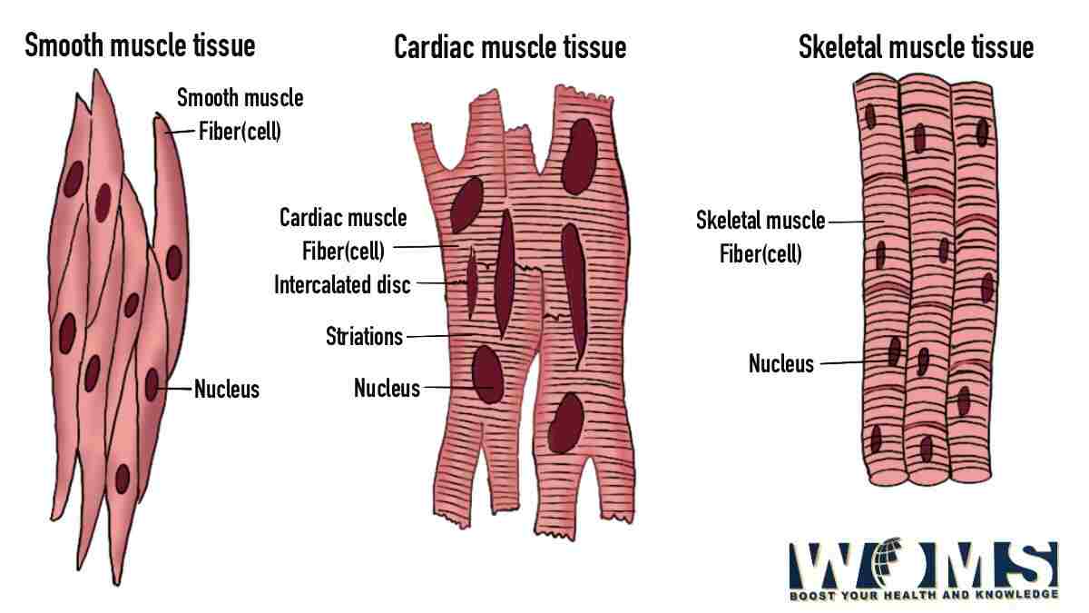
Our body possesses three main kinds of muscles that perform most of the body’s functions. These are as follows:
- Skeletal muscle
- Smooth muscle
- Cardiac muscle
These muscles contain different types of muscle fibers performing multiple functions. Let us have a detailed look at these muscles concerning their diagram of muscle fibers. Every muscle possesses a different muscle fiber type performing several functions.
Cardiac muscle fiber
Cardiac muscles are the primary muscles associated with the heart myocardium. These muscles help pump blood outside the heart through organized contractions. Cardiac muscle also contains muscle fibers to perform their normal function. Cardiac muscle fibers show some similarities to skeletal muscle fibers. But there is some difference that creates a proper boundary line between these two muscle fibers.
Cardiac muscle fibers contain syncytium with a proper connection through intercalated discs. This can be demonstrated in the diagram of muscle fiber. These intercalated discs join one cardiomyocyte to the other associated cardiac muscle fibers. In this way, it offers structural stability to the myocardium of the heart. In addition, these junctions help to transmit the continuous flow of action potential throughout this muscular layer. Moreover, cardiac muscle fibers are striated due to the presence of the A and I bands.
The striated orientation of cardiac muscle fibers dictates the smooth layers of actin and myosin filaments. These striations can be demonstrated in the diagram of muscle fiber. In addition, cardiac muscle fibers also contain sarcoplasm and several other organelles to maintain the proper appearance of a cell. There are multiple abundant myofibrils arranged in a parallel direction. These are the main contractile proteins. With the help of these structures, cardiac muscles work properly to contract the heart and pump blood outside the aorta. You can relate various structures in the diagram of muscle fiber.
Smooth muscle fiber
Smooth muscle fibers are not striated as seen in the diagram of muscle fiber. They offer quite a smooth appearance without any presence of any bands. Smooth muscle fibers exhibit an oblong to spindle shape appearance wider from the middle and tapered toward the ends. The smooth muscle fibers are way shorter than the striated muscle types. They don’t contain striation but contain actin and myosin for the contraction.
Skeletal muscle fiber
Skeletal muscle contains thousands of muscle fibers tightly connected to each other through connective tissue. Every muscle fiber contains repeating units of thick and thin filaments. These thick and thin filaments give a proper striated appearance to the skeletal muscle. There are three types of muscle fibers in skeletal muscle. Skeletal muscle fibers can be demonstrated in the diagram of muscle fiber. These are as follows:
- Type 1 – Slow oxidative
- Type 2A – Fast oxidative
- Type 2B – Fast glycolytic
Skeletal muscle contains all these muscle fibers in different proportions. Moreover, these muscle fibers can adapt themselves according to the body’s demands. They adapt themselves by changing their size and composition. Does this mean they can also help you get bigger hands? Let us have a detailed look at the different types of these muscle fibers to understand the details of the diagram of muscle fibers.
Type 1 – Slow oxidative muscle fibers:
As the name indicates, these muscle fibers contract more slowly than the other muscle fiber types. In addition, these muscle fibers utilize oxygen and glucose (aerobic respiration) to overcome energy demands through ATP. Type 1 fibers contain a higher amount of energy powerhouses in the form of mitochondria. These organelles give a darker appearance to the s fibers.
These muscle fibers offer low-power muscle contractions. But these slow contractions are for longer periods and offer more resistance to fatigue. Fatigue resistance makes these muscle fibers more useful in maintaining balanced muscular contractions, posture maintenance, making small movements, and stabilizing the movements of bones and joints.
These movements occur more often but utilize a very low quotient of energy. These muscle fibers are not used for more powerful and energy-demanding workouts. Moreover, this muscle fiber type contains high myoglobin content. Therefore, these fibers are also known as red fibers as also seen in the diagram of muscle fiber.
Type 2A – Fast oxidative muscle fibers
These muscle fibers also utilize glucose and oxygen just like the slow oxidative muscle fiber type. These muscle fibers are also known as intermediate fibers because of their features lying intermediate between the fast and slow twitch fibers. As these muscle fibers are quite fast, they produce ATP faster than the slow twitch fibers. They contain relatively fewer mitochondria than the slow oxidative muscle fibers. These structures can be observed in the detailed diagram of muscle fibers.
This muscle fiber type contains less myoglobin than the slow oxidative muscle fibers. Therefore, these fibers are lighter than the slow oxidative muscle fibers. These muscle fibers also show fatigue resistance. Moreover, these fast oxidative muscle fiber types require more energy than the slow twitch to work smoothly. These muscle fibers are useful for sprinting type of movements because of increased tension production.
Type 2B – Fast glycolytic muscle fibers
These muscle fibers do not utilize the aerobic mode of respiration. Instead of using oxygen and glucose, these muscle fibers store energy to be used for hectic movements. In addition, these muscle fibers utilize the method of anaerobic glycolysis to produce ATPs as an energy source.
These muscle fibers are larger in diameter as seen in the diagram of muscle fiber with more amounts of glycogen to produce energy. Moreover, this muscle fiber type does not possess a higher density of mitochondria and myoglobin. Therefore, these muscle fibers are whiter than the previous muscle fiber types.
Besides these characteristic features, these muscle fibers are not fatigue resistant. That’s why these fibers offer high-energy movements for a very small time. These fast glycolytic muscle fibers are used for powerful but rapid movements.
| Characteristic features: | Type 1- Slow oxidative muscle fiber | Type 2A – Fast oxidative muscle fiber | Type 2B – Fast glycolytic muscle fiber |
| Activities: | Posture maintenance, isometric contraction, making small movements | Sprinting, jumping, powerlifting | Rapid movements |
| Diameter of muscle fiber | Smaller in size | Larger | Larger |
| Fatigue resistance | Slow | Quickly resistant | Very quickly resistant |
| Force production | Low | High | Very high |
| Speed of contraction | Slow | Quick | Very quickly |
| Mitochondria amount | High-density | Medium | Low-density |
| Capillaries | High amount | Medium | Low amount |
| Myoglobin | High amount | Medium | Low amount |
| ATPase level | Low-level | Medium level | High-level |
Clinical conditions associated with muscle fibers
After knowing the structures of muscle fibers by looking at the diagram of muscle fiber, we now move into the clinical aspects. Muscle fibers are more prone to suffer from various medical problems. There is a list of problems associated with muscle fibers. Some of the common disorders of muscle fibers are as follows:
Muscle cramps
This problem arises when a single muscle fiber or a group of muscles contracts involuntarily. It is quite a painful condition that may last for seconds or even minutes. This condition heals itself after some time.
Muscle palsy
This is the problem arising due to any problem in the motor function of the nerves innervating the muscles. In this condition, a partial or complete loss of muscle function may occur. This condition may lead to weakness or paralysis of the muscles. The most common diseases associated with muscle palsy are Guyon canal syndrome and Bell’s palsy.
Muscle trauma or injury
This condition occurs when muscle fibers get stretched or injured due to any traumatic movement. It usually occurs when muscles stretch beyond their normal limit or move forcefully. These kinds of injuries are common during sports or accidents.
Asthma
Asthma is a respiratory problem arising due to the sudden contraction of a smooth muscle when any stimulus hits the respiratory tract. This condition can cause sudden narrowing of the airway tract leading to different breathing problems.
Coronary artery disease
This condition is associated with cardiac muscle fibers. It occurs when there is a lack of oxygen to cardiac muscles leading to the symptoms of angina. This may cause damage to the cardiac muscle fibers leading to the dysfunction of the heart. You should follow healthy heart tips to keep your heart healthy.
Atrophy of the muscle
It is a condition related to the degeneration of the muscle fibers. Moreover, this condition may cause the loss of muscle volume with weakness.
Conclusion
Every muscle contains its muscle fibers. There are different types of muscle fibers depending on the muscle type that can be seen in the diagram of muscle fiber. Cardiac muscle fibers offer a striated appearance and work to contract the heart. Whereas, smooth muscles offer a smooth appearance with no striations. In contrast to smooth muscle, skeletal muscle fibers are striated and of three types. These are slow oxidative, fast oxidative, and fast glycolytic muscle fibers.
These muscle fibers perform their function accordingly. Moreover, these muscle fibers can also cause medical problems. You may consult your physician regarding the health of your muscles.
Frequently asked questions (FAQs)
What are the common problems associated with aging?
There are several problems associated with the aging process. The most common problems are the atrophy of the muscles and degeneration of the nerve fibers leading to the loss of function.
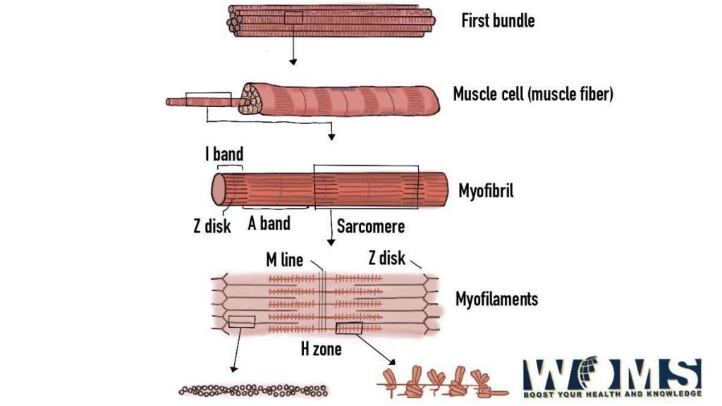
What is the composition of the muscle fiber?
Muscle fibers are made up of myofibrils. These myofibrils contain actin and myosin as contractile proteins to perform the main function of the muscle.