Which is the longest bone in human body?
The longest bone in human body
Since childhood, you may have also learned that there are 206 bones in an adult human body. But did you ever wonder which among these is the longest bone in the human body?
As many of you already know, it’s Femur. Also called the thigh bone, Femur is the longest bone in the human body. As bones provide us strength, Femur being the longest bone is also the strongest bone in
human body.
While talking about the longest bone, some of you may also be wondering of the shortest one. The shortest bone in the human body is stapes.
Now let’s return to our topic of the longest bone of the human body. You may be surprised to know the length of Femur. Femur measures are almost 1/4th of the height of a normal adult human. In numbers, it measures almost 19.9 inches. A detailed description of the longest bone in the human body will be included in this article.
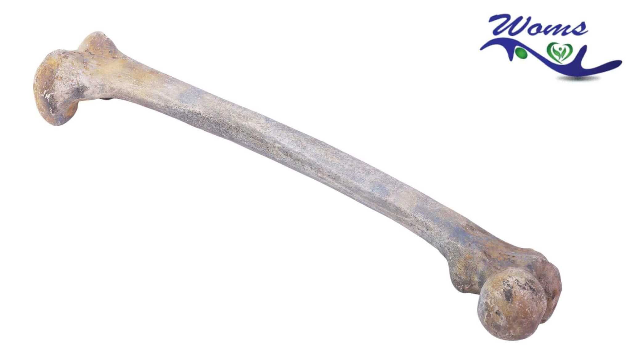
The anatomical position of Femur
The head portion of Femur remains medially directed upwards while slightly leaning forward. Similarly to keep the lower surfaces of the two condyles lie in the same plane, the shaft of Femur is directed medially and obliquely downwards.
Side determination of the longest bone in the human body
– There is a round head in the upper end of the longest bone in the human body.
Similarly, the lower end is wide forming two large condyles
- The upper end of the head of the femur medially directed
- The shaft follows convex way forward
Features of the longest bone of the human body
The features of each portion of Femur are mentioned below:
Head of Femur
In the upper end of Femur, there is the presence of a head which is like half a sphere and is medially directed upwards being slightly forwarded. The neck, intertrochanteric crest along with the greater trochanter is commonly described as the head of Femur.
The head part further articulates with the acetabulum forming the hip joint. There is a pit present behind the centre and just below the head called Fovea.
Neck of Femur
Measuring about 3.7 centimetres in length, the neck of the longest bone of human body makes the connection between the head and the shaft.
There are two surfaces in the neck of Femur, the anterior surface and the posterior surface.
The anterior surface of the neck of Femur meets the shaft near the intertrochanteric line and is flat. This is the surface where the articular cartilage of the head may extend up to.
The posterior surface of Femur seems concave from side to side while convex from above and downwards. The posterior surface meets the shaft at the intertrochanteric crest. The anterior surface of Femur is completely intracapsular while in the posterior surface, just little more than it’s medial half is intracapsular.
Similarly, there are two borders in the neck of the longest bone. They are the upper border and the lower border.
The upper border connects to the shaft at the greater trochanter. The upper border is concave and horizontal.
The lower border meets the shaft at the less trochanter and is oblique and straight.
Greater trochanter
In the upper part of the neck-shaft junction, there is the presence of abundant quadrangular structure called the greater trochanter.
The greater trochanter of the longest bone in human body consists of three surfaces along with an apex. The three surfaces are anterior, medial as well as lateral.
Lesser trochanter
At the junction of a posteroinferior portion of the neck and shaft, there is the presence of a conical structure which is medially directed and lies backwards of the junction.
Intertrochanteric line
There is a line that marks the point of a function of the anterior portion of the neck with the shaft. This is called the intertrochanteric line.
Intertrochanteric crest
There is a smooth and round ridge-like structure that marks the point of junction of the poster part of the neck with the shaft of the longest bone of human body. This ridge-like structure is called the intertrochanteric crest.
The shaft
The shaft of Femur is a cylindrical structure which is narrow in the middle while expanded more inferiorly than superiorly.
Looking from forward, the shaft looks convex and directed medially and obliquely downwards.
The lower end of Femur
As mentioned above, the lower end of the longest bone in the human body is wide and expanded to form two condyles.
Among the two large condyles, one is medial while the other is lateral.
Both the condyles are anteriorly united. Anteriorly, they are in line with the front part of the shaft. While talking about the posterior side, the condyles are separated by a deep gap. This deep gap is named as an intercondylar notch or intercondylar fossa.
Articular surface
There is a surface by which the condyles of the longest and strongest bone of the human body is partially covered. This surface is known as the articular surface. Tibial and patellar parts are the two parts of the articular surface.
The patellar part of the articular surface covers the anterior portion of the condyles. The extension of the patellar part is more on the lateral condyle than on the medial condyle.
In the same way, the tibial part of the articular surface covers the posterior and inferior portions of the condyles.
The tibial surface also merges with the patellar surface anteriorly.
Lateral condyle
Among the two condyles, this is a laterally flat condyle and is in line with the shaft of Femur. The lateral condyle is highly responsible for the transmission of body weight to the tibia.
The lateral condyle is less prominent but stronger and stouter than the medial condyle.
Medial condyle
Another condyle is the medial condyle that appears medially convex. The medial epicondyle is the most prominent part of the medial condyle. In the posterior region, superior to the epicondyle, a projection called adductor tubercle is present. This is an important landmark as for the lower end of Femur, the epiphyseal line
passes through it.
Intercondylar fossa
Also called, Intercondylar notch, this is a notch that separates the posterior as well as lower parts of the condyles. The intercondylar fossa is anteriorly limited by the patellar part while posteriorly by the Intercondylar line. The notch is separated from the popliteal surface by the Intercondylar line.
The nutrient artery of the longest bone of human After hearing so many terms about the strongest bone of the human body, you may be wondering about the nutrient artery of Femur.
The nutrient artery of Femur is the second perforating artery. It is one of the branches of the profundal femoris artery. Location of the nutrient artery Regarding the location of the artery, it is situated near the medial side of linea apsera. The artery is upwardly directed.
FAQs:
1. Which is the shortest bone of our body
Stapes is the shortest bone of our body as it is 3 x 5 mm in size.
2. Which bone is known as the beauty bone?
The clavicle bone is known as the beauty bone of the body.
3. Which is the softest bone in the body?
Clavicle bone is the softest and weakest bone in the body
4. Which is the weakest muscle present in our body?
Stapedius muscle is the weakest muscle present in our body.
5. Which is the strongest muscle in our body?
The muscle which is strongest in our body is the muscle Masseter as it forms the muscle of jaw.