Pharynx Anatomy: Muscles, Blood Supply and Innervations
In terms of pharynx anatomy, it is the semicircular fibromuscular tube-like structure that is usually known as the throat in layman’s language. The pharynx is located anteriorly to the vertebral column. In addition, the pharynx boundaries start from the base of the skull and extend up to the lower border of the cricoid cartilage. It is about 5cm in length. This article completely covers the diverse details of pharynx anatomy to learn the basic concepts. Let us have a look at the anatomical aspects of the pharynx concerning the pharynx anatomy.
Gross pharynx anatomy
Understanding pharynx anatomy is divided into two sections.
- External section – it includes posterior and lateral views including the external surface of muscles that forms the wall of the pharynx.
- The internal section – includes a sagittal view to describe the pharynx anatomy.
The external surface of the pharynx anatomy
Considering the posterior aspect, the pharynx lies anterior to the cervical vertebrae exteriorly. From the upper limit, it communicates with the occipital bone anteriorly to the foramen magnum. The external surface of muscles includes voluntary muscles which are covered by buccopharyngeal fascia externally. There are four primary muscles associated with the external surface of the pharynx. These are as follows:
Superior pharyngeal constrictor – This muscle has its origin in different anatomical structures of the lateral face. These structures are the posterior border of the mandibular raphe, pterygoid hamulus, posterior aspect of the mylohyoid sling of the mandible, and the lateral side of the tongue. Connecting all these structures, the fibers of this muscle terminate at the pharyngeal tubercle located on the basilar portion of the occipital bone. This is an important muscle of pharynx anatomy.

- Middle pharyngeal constrictor – This muscle lies below the level of the superior pharyngeal constrictor muscle. This muscle has its origin in the stylohyoid ligament with Greater and Lesser horns of the hyoid bone. Some of the fibers of these muscles terminate by blending with the superior constrictor muscle. Whereas, the others insert into the inferior pharyngeal constrictor muscle by overlapping the fibers.
- Inferior pharyngeal constrictor – This muscle is split into two distinct muscle groups.
- Cricopharyngeus – had its origin from the cricoid cartilage. Whereas, in contrast to other muscles of the pharynx, it terminates into the circular muscles of the esophagus.
- Thyropharngeus – originates from the thyroid cartilage and terminates at the pharyngeal raphe.
- Stylopharyngeus – This muscle has its origin in the medial aspect of the styloid process of the temporal bone. The fibers of this muscle terminate by blending with the fibers of superior and middle constrictor muscles. Some leftover fibers have an insertion point lying on the posterior surface of the thyroid cartilage.
In addition, there are also longitudinal muscles associated with the external surface of the pharynx. These are as follows:
- Palatopharyngeus – This muscle has an origin on the posterior aspect of the hard palate at the level of the palatine aponeurosis. Whereas, it terminates by blending with the opposite side of the palatopharyngeus muscle at the posterior boundary of the thyroid cartilage.
- Salpingopharyngeus – This muscle has its origin in the cartilaginous portion of the auditory tube and terminates by blending into the palatopharyngeus muscle
The function of the pharynx
After knowing the pharynx anatomy, one can easily guess the function of pharynx. Functions of the pharynx are quite obvious to understand and learn.
- The tubular surface of the pharynx allows the air, food, and other liquids to pass from the mouth to the lower GI tract.
- It helps in both the digestive functions as well as respiratory functions of the human body.
- Muscles of the pharynx help in the peristalsis movement along with speaking and swallowing.
Associated structures with the external surface of the pharynx:
There are several important structures lying in association with the external surface of the pharynx. It is important to know these structures while knowing about pharynx anatomy. These structures are as follows:
- Structure lying superiorly and posterolaterally to the pharyngobasilar fascia – jugular foramen serving as a leaving site for the cranial nerves IX, X, and XI. Cranial nerve IX leaves the jugular foramen anteriorly to the other two cranial nerves.
- Posterior the cranial nerve IX, X, and XI – internal jugular vein leaves the jugular fossa
- Medially to the jugular foramen – cranial nerve XII leaves the hypoglossal canal
- The gap presents superiorly to the superior pharyngeal constrictor muscle – This point serves as an entry point for the auditory tube and levator veli palatini muscle.
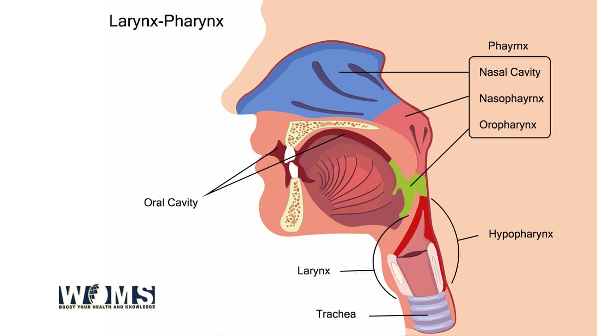
Internal surface of the pharynx anatomy:
The internal surface of the pharynx is divided into three main portions to understand pharynx anatomy thoroughly. These are as follows:
- Nasopharynx – it starts from the region superior to the soft palate that connects the nasal cavity. Its inferior border is the pharyngeal isthmus. From this point, the oropharynx starts.
- Oropharynx – it is the medial portion of the pharynx forming a communication between the oral cavity and pharyngeal isthmus.
- Laryngopharynx – it is the last portion of the pharynx. On its superior aspect, there is epiglottis or oropharynx. Whereas, at its inferior border, there is the posterior aspect of the cricoid cartilage of the larynx.
Blood supply to the pharynx
The pharynx receives its sufficient blood supply through various arteries. Moreover, it also gets enough blood supply through the arterial anastomosis. These features make the pharynx one of the richest structures getting enough blood supply. All of the arterial blood supply originates from the external carotid artery. There are some of the common arteries of the pharynx, providing blood to the muscles of the pharynx.
- Maxillary artery
- Lingual artery
- Facial artery
| Muscles of the pharynx: | Blood supply to these muscles: |
| Superior pharyngeal constrictor: | Pharyngeal branch of the ascending pharyngeal artery Tonsillar branch of the facial artery |
| Middle pharyngeal constrictor: | Pharyngeal branch of the ascending pharyngeal artery Tonsillar branch of the facial artery |
| Inferior pharyngeal constrictor: | Pharyngeal branch of the ascending pharyngeal artery Muscular branches of the inferior thyroid artery |
| Palatopharyngeus: | Ascending palatine branch of the facial artery descending palatine branch of the maxillary artery Pharyngeal branch of the ascending pharyngeal artery |
| Salpingopharyngeus: | Ascending palatine branch of the facial artery descending palatine branch of the maxillary artery Pharyngeal branch of the ascending pharyngeal artery (similar to the Palatopharyngeus muscle) |
| Stylopharyngeus: | Pharyngeal branch of the ascending pharyngeal artery |
In addition, these pharyngeal muscles drain their deoxygenated blood through the external palatine vein. This major vein drains into the pharyngeal plexus and ultimately into the internal jugular vein. The internal jugular vein and anterior jugular vein are different. The blood supply of pharynx is a must know thing while reading about pharynx anatomy.
Innervation to the pharynx
Pharynx except the nasopharynx drives its sensory nerve supply from the glossopharyngeal nerve. More commonly, it gets its nerve supply from the pharyngeal and tonsillar branches of the glossopharyngeal nerve. The nasopharynx drives its nerve supply from the branch of the maxillary nerve, known as the pharyngeal nerve.
The pharyngeal branch of the vagus nerve offers motor innervation to all muscles except the stylopharyngeus and other related structures of the pharynx. Stylopharyngeus receives its motor supply through the glossopharyngeal nerve through fibers of the nucleus ambiguous.
Summarizing the details related to the innervation of the pharynx anatomy, there are three main nerves providing the sensations and functional stability to the muscles of the pharynx. These are as follows:
- Glossopharyngeal nerve – sensory innervation except for Nasopharynx
- Maxillary nerve – sensory innervation to Nasopharynx
- Vagus nerve – motor innervation of all muscles of the pharynx except the Stylopharyngeus
- Stylopharyngeus receives motor supply through the glossopharyngeal nerve
Clinical conditions associated with the pharynx anatomy
The pharynx is the main structure in the neck region performing various functions of daily life. Any changes or disturbances in the anatomical features of the pharynx or the pharynx anatomy may lead to dysfunction. Some of those medical conditions are as follows:
Pharyngitis
It is a disease associated with the inflammation of the pharynx also known as a sore throat. There are several agents that may cause inflammation of the pharynx. Viruses are the most common leading agents. The most common symptoms are pain, irritation, discomfort, and difficulty in swallowing.
Adenoiditis
There are several adenoids present in the surrounding of the pharynx to drain lymph fluid from the various structures. It usually occurs due to the presence of any infection. It usually exhibits flu-like symptoms. Most commonly, it includes nasal obstruction, rhinorrhea, and breathing issues.
Obstructive sleep apnea
Sleep apnea may happen due to the larger lymph nodes or excess of the soft palate. But, it can also happen due to oropharyngeal collapse. The muscles of the pharynx may become hypotonic, leading to difficulty in breathing.
Cricopharyngeal Achalasia
It is a rare disease occurring due to the weakness of the cricopharyngeus. When bolus passes from the esophagus, it tends to leak making it difficult to eat and swallow.
Frequently asked questions (FAQs):
What are the common structures present within the boundaries of the pharynx?
While reading pharynx anatomy, we see the pharynx contains the following structures:
● Three sets of tonsils which are located on the posterior surface of the throat and at the base of the tongue. These are helpful in the defense mechanism of the body.
● Eustachian tubes which are also known as auditory tubes. These tubes interlink the ears to the pharynx. Moreover, these are helpful to maintain pressure and drain the fluids.
When there is a need to contact a doctor regarding the medical conditions of the pharynx?
If the symptoms of your disease persist, you must contact your doctor immediately. The symptoms must include
● Pain in ear
● High grade fever indicating infection
● Any lump in the neck or throat region
● Sore throat continuing for more than 5 to 7 days
● Difficulty in swallowing
● Stiffness in the neck