The ultimate guide to Pig skull: Images and Anatomy
Are you searching for anatomical landmarks of the pig skull? There is no need to scroll multiple websites to compile all the details related to it’s anatomy. This article provides a clear anatomical illustration related to the pig skull.
Veterinary students are curious about the easy and precise anatomical details related to animals. Pigs are important due to some osteological differences between wild and domestic pigs. Let us provide a clear perception regarding the differences and concepts related to their skull. Continue reading the article and build your concepts based on these anatomical details.
What are the important anatomical features of the pig skull?
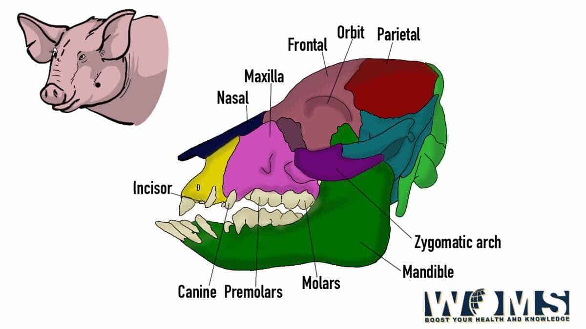
The pig skull is usually elongated with multiple variations in shape and length depending on the pig species. The facial part of the skull is generally elongated. Moreover, the frontal aspect of the pig skull is usually straight. The brain is present within the cranium. The cranium is the posterior portion of the skull. The brain maintains its connection to the spinal cord through the foramen magnum.
There are different bones, either paired or unpaired، present in the pig skull connected through different sutures. The pig skull is quite distinctive in shape with a sloping crest to the back. The pig skull is usually formed by the supraoccipital and different other important bones. In addition, there is also a pre-nasal bone present at the prominent tip of the snout.
What are the different bones present in the pig skull?
There are different bones that form the pig skull. Let us have a look at the anatomical details of the bones, forming their skull.
The occipital bone of the pig skull is usually flat and elongated, making the caudal portion. It ensures the complete formation of the nuchal walls and foramen magnum. Moreover, the bacillary aspect of the occipital bone is hexagonal, rostrally thicker, and caudally flattened. It contains muscular tubercles on its ventral aspect to which flexors of the head and neck are attached. In addition, the squamous aspect of the occipital bone is quite extensive, flattened, and triangular.
The nuchal crests serve as a landmark for the collection of cerebrospinal fluid. There are also occipital condyles that show articulation with the atlas, forming atlanto occipital joints. This orientation of the occipital bone offers a definitive shape to their skull.
The sphenoid bone of the pig skull contains the body and pairs of wings. It forms the basal surface of the neurocranium. These bones are separated through cartilage. Cartilages continue to ossify with age. There are two parts of the sphenoid bone.
- The presphenoid is present rostrally and contains a caudal fossa with sphenoid sinuses present on the inner surfaces. These sinuses contain an optic canal and optic chiasma.
- On the caudal aspect, there is a basisphenoid with median cranial fossa.
The wings of the sphenoid bone lie opposite to the temporal bone, orbit, maxilla, and brain. In addition, these wings also form foramen ovale and different other foramina. Moreover, the pterygoid processes are also available.
The frontal bone is a paired bone connected through the interfrontal suture present between the cranium and face. It also encloses the frontal sinuses. The nasal and lacrimal bones are present at the borderline of the frontal bone
The temporal bone consists of three main parts known as squamous, tympanic, and petrosal. These parts maintain their communication and firm the lateral wall of the pig skull. In addition, the temporal bone articulates with the frontal, sphenoid, and parietal bones.
- The squamous parts connect the temporal process of the zygomatic bone to the zygomatic arch. In this way, it forms the articulating surfaces of the temporomandibular joint.
- The tympanic part is present on the ventral aspect of the temporal bone, containing the tympanic bulla. In addition, the tympanic membrane differentiates the tympanic cavity from 5he external acoustic meatus. Moreover, it also surrounds the auditory ossicles dorsally.
- The petrosal part of the temporal bone surrounds the inner ear internally through the internal acoustic meatus. In the ventral direction, it forms the mastoid process.
The parietal bone is also a paired bone, forming the dorsolateral wall of their skull. It is made up of the parietal plane, nuchal plane, and temporal plane. There is also an interparietal bone present between the parietal and occipital bone. It fuses with age.
The ethmoid bone is present in deeper portions of the pig skull. It is present in between the facial and cranial portions of the pig skull. In addition, it is separated from the cranial cavity through the cribriform plate. There are several small foramina in which olfactory nerves exist.
The nasal bone is a paired bone, forming the bridge of the nasal cavity. The ethmoidal crest is connected internally to the dorsal nasal conchae. A rostral suture,nasoincisive notch, is present on the apex between the incisive and nasal bone. In a similar way, you can have a look at the various bones present in The Lion skull.
The lacrimal bone is present on the rostral margin of the orbital bone. Lacrimal bone is quite irregular in shape. It forms the lateral wall of the face and orbit, present near the medial canthus. In addition, it also maintains its connection with frontal, zygomatic, and maxillary bones.
The zygomatic arch or zygomatic bone is stronger and flattened from one side to the other side. It is present laterally and ventrally to the lacrimal bone. The facial crest is present on the lateral aspect of the zygomatic bone. Skull of pig also contains an incomplete or small orbital cavity to adjust the eye content.
The incisive bone is a paired bone, made up of body, palatine, nasal and alvrolar parts. It communicates with the maxilla to firm the interalveolar margin. In addition, it also forms the rostral portion of the facial part of the pig skull.
The palatine bone is also a paired bone located between the maxilla, pterygoid, and sphenoid bones. It is made of horizontal plates evolving to form a hard palate. Moreover, it also contains a perpendicular plate and choanae. The horizontal plane also contains a palatine sinus.
The vomer is an unpaired bone, extending from the choanae to the floor of the nasal cavity. In addition, it also connects the median nasal crest and possesses a septal sulcus surrounding the nasal cavity.
The pterygoid bone is a paired bone limited by the palatine and sphenoid bones. It makes the dorsolateral wall of the nasopharyngeal cavity.
The maxilla, the key bone of the upper jaw, contains the upper dentition. It contains maxillary sinuses forming the external orientation of the face. In addition, it also forms the facial crest.
The mandible is divided into the ramus and the body. The body supports the incisor teeth in the rostral direction and cheek teeth in the caudal direction. The mandible also contains mandible canal and mental foramen.
How are the bones connected in the pig skull?
There are different bones in their skull that are connected with each other. Sutures are the main connecting points that offer continuity between the bones. Sutures joints are usually in a jigsaw pattern engraved on the surface of their skull. These sutural joints indicate that the skull is formed by the combination of several bones. The skull joints are usually immovable, restricting every kind of movement.
A saw-cut pattern passes transversely from the facial aspect of the skull. This pattern indicates the presence of turbinate bone in the nasal cavity. These turbinate bones provide a supportive role for the nasal epithelium to moisten and warm the air moving from outside toward the lungs. In addition, it offers a larger surface area for the sensation of smell.
What is the main function of the pig skull?
The pig skull offers a variety of functions including:
- Protecting the brain
- Foramen development for the exit and entry of various nervous and defensive structures
- Support system for the facial muscles to offer origin and insertion sites
What are the major foramen and canals present in the pig skull?
There are various structures associated with the pig skull. Skulls provide a protective barrier for a variety of structures. These are as follows:
- Foramen magnum – passing site for the spinal cord. In addition, the alar ligament also passes through the foramen magnum along with vertebral arteries.
- Jugular foramen – it contains the internal carotid artery, glossopharyngeal nerve, accessory nerve, and vagus nerve. In addition, it is present adjacent to the tympanic bulla.
- Stylomastoid foramen – it allows the facial nerve to pass and present on the petrosal aspect of the temporal bone.
- Ethmoidal foramen – it allows the ethmoidal artery, vein, and olfactory nerve to pass.
- Mental foramen – it allows the mental artery and vein to pass along with the mandibular branch of the trigeminal nerve.
Conclusion:
A pig skull contains a variety of bones, with every bone having its own anatomical importance. These bones are associated to maintain the continuity of the skull. The skull encloses the brains and different other structures. In addition, there are several muscles associated with the pig skull to orient different kinds of movements. This article explained the anatomical concepts related to the pig skull and its associated structures. Hope you will be able to correlate this knowledge to your clinical concepts.
Frequently asked questions (FAQs)
Which kind of skeleton do pigs have?
Pigs possess two types of skeleton including both the axial and appendicular skeleton.
How many teeth are present in the pig skull?
Pigs usually possess 44 teeth in both upper and lower arches. There are three incisors, one canine, four premolars, and three molars for every quadrant.