Middle meningeal artery
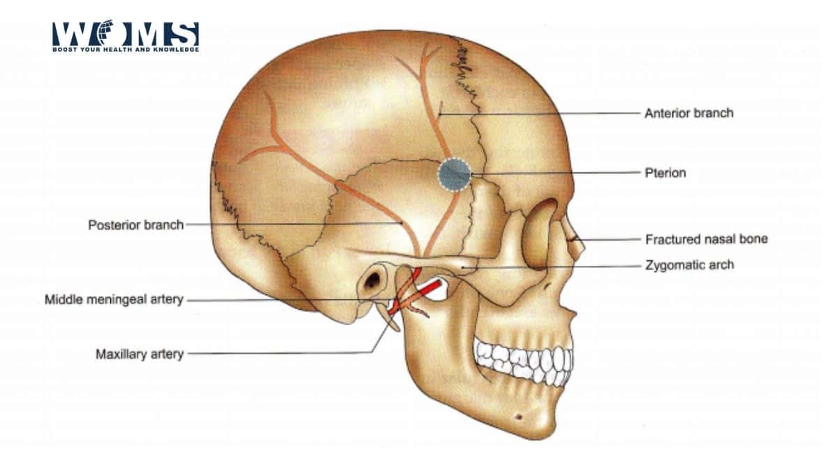
The middle meningeal artery is the largest of the meningeal arteries. It arises from the first part of the maxillary artery in the infratemporal fossa and passes between the roots of the auriculotemporal nerve. It lies lateral to tensor Veli palatine, then enters the cranial cavity through the foramen Spinosum.
The middle meningeal artery is a vital artery of the brain that has an important clinical role. It is important to the surgeon because this artery is the commonest source of extradural haemorrhage, which is an acute surgical emergency.
The middle meningeal artery (MMA) normally arises from the maxillary artery, a terminal branch of the external carotid artery. It runs through the foramen Spinosum to supply the dura mater (the outer meningeal layer), and the calvaria. It is the largest of three (paired) arteries that supply the anterior meninges, the other being the anterior meningeal artery and the posterior meningeal artery.
Dissection
You need to dissect the middle meningeal artery which enters the skull through foramen Spinosum. It is an important artery for the supply of endocranium, inner table of skull and dipole. You need to examine the other structures seen in cranial fossae after the removal of the brain. These are the cranial nerves, internal carotid artery, petrosal nerves and fourth part of the vertebral artery.
Origin
The Middle meningeal artery is a branch of the first part of the maxillary artery ( which is a branch of ECA) given off in the infratemporal fossa.
Branches of the middle meningeal artery
In middle cranial fossa, it divides into frontal and parietal branches.
Terminal branches
- Ganglionic branches-to-trigeminal ganglion.
- A petrosal branch-to facial nerve, ganglion, and tympanic cavity.
- A superior tympanic artery-to tensor tympani muscles.
- Temporal branches-to temporal fossa
- An anastomotic branch—anastomoses with the recurrent branch of the lacrimal artery.
Course and relations
- In the infratemporal fossa, the artery runs upwards and medially deep to the lateral pterygoid muscle and superficial to the sphenomandibular ligament. Here it passes through a loop formed by the two roots of the auriculotemporal nerve.
- It enters the middle cranial fossa through the foramen Spinosum.
- In the middle cranial fossa, the artery has an extradural course, but the middle meningeal veins are closer to the bone than the artery. Here the artery runs forwards and laterally for a variable distance, grooving the squamous temporal bone, and divides into a frontal and parietal branch.
- The frontal or anterior branch is larger than the parietal branch. First, it ambles forwards and laterally towards the lateral end of the lesser wing of the sphenoid. Then it runs obliquely upwards and backward, parallel to, and a little in front of the central sulcus of the cerebral hemisphere. Thus after grating the pterion, the artery is closely related to the motor area of the cerebral cortex.
- The parietal or posterior branch runs backwards over, or near, the superior temporal sulcus of the cerebrum, about 4 cm above the level of the zygomatic arch. It ends in front of the posteroinferior angle of the parietal bone by dividing it into branches.
Importance
It is of great surgical importance because its tear in head injuries may cause extradural haemorrhage. Rupture of the middle meningeal artery most commonly results in an epidural (extradural) hematoma, a neurosurgical emergency that may present with a lucid interval followed by rapid neurological deterioration. The hematoma presses on the motor area, giving rise to hemiplegia of the opposite side. The anterior division can be approached surgically by making a hole in the skull over the pterion, 4cm above the midpoint of the zygomatic arch.
Rarely the parietal branch is implicated, causing contralateral deafness. In this case, the hole is made at point 4 cm above and 4 cm behind the external acoustic meatus.