Femoral Vein Anatomy: Tributaries and 3 Clinical Significance
The femoral vein is the largest blood vessel located deep in the upper thigh region thus learning femoral vein anatomy becomes important. The femoral vein moves along the femoral artery in close proximity. It collects deoxygenated blood from different tissues of the lower leg and takes it toward the heart, converting it to oxygenated blood. Once the blood becomes oxygenated, it moves back towards the lower leg through the femoral artery.
This article covers the details of femoral vein anatomy in context to the origin, course, tributaries, and other specific features. Let us take a deep dive into the anatomical aspects of the femoral vein to understand the concepts of femoral vein anatomy.
Gross femoral vein anatomy
The femoral vein is one of the largest blood vessels with a diameter of 12 to 14 millimeters in adults, equal to half an inch. It continues originating near the knee and grows in size moving towards the upper thigh. Just like other blood vessels of the human body, the femoral vein also consists of three main cell layers. Let us learn femoral vein anatomy in details. These layers are as follows:
- Tunica Intima – It is the inner lining of the femoral vein containing squamous cell epithelium. In addition, it also includes connective tissue with a semipermeable cell layer.
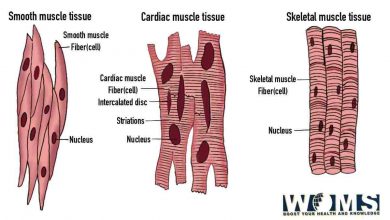
- Tunica Media – It lies intermediate between the inner and outer lining of the cell layer. It is a relatively thicker cell layer containing smooth muscle. The smooth muscle helps to apply pressure to push the blood along the vein and opposite to gravity.
- Tunica Extrema – It is the outer cell layer with different amounts of rigid and elastic fibers. These fibers are helpful for the femoral vein to maintain its shape and position.
Course and Origin of the femoral vein
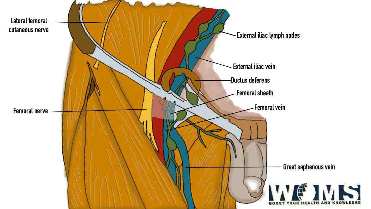
The femoral vein originates from the continuation of the popliteal vein and passes through the adductor hiatus. The popliteal vein drives deoxygenated blood from the tibial veins of the lower leg. In this way, the popliteal vein continues to produce the femoral vein. Moreover, the femoral vein continues to move towards the anterior aspect of the thigh. It extends further upward and towards the body’s center direction along the adductor canal. Along the course, the femoral vein also gets access to the femoral triangle (triangular depression between the muscles of the thigh).
The femoral vein also crosses the femoral sheath. At this point, just behind the inguinal ligament, the femoral vein continues into the external iliac vein. Moreover, mentioning in the femoral vein anatomy, it terminates at this region, draining the blood into the external iliac vein. This was about the origin and course of femoral vein anatomy. You can similarly have a look at the veins of upper limb.
The function of the femoral vein
All veins other than the pulmonary vein carry deoxygenated blood from the associated tissues toward the right side of the lung. The heart takes this blood and transports it to the lungs through the pulmonary artery. Lungs convert the deoxygenated blood to oxygenated blood. Then, pulmonary veins take back this oxygenated blood to the left side of the heart. The heart pumps this blood to all body tissues and organs through the aorta.
Concerning the femoral vein anatomy, it drains blood from the lower limb of the human body. The popliteal vein collects blood from the feet and back of the lower leg and continues ad femoral vein at the level of the knees. Moving towards the center of the body, the femoral vein also drains blood from the associated muscles of the thigh.
Tributaries of the femoral vein:
There are multiple tributaries that continue to drain blood into the femoral vein, as it moves in an upward direction toward the body’s center. Some of them are as follows:
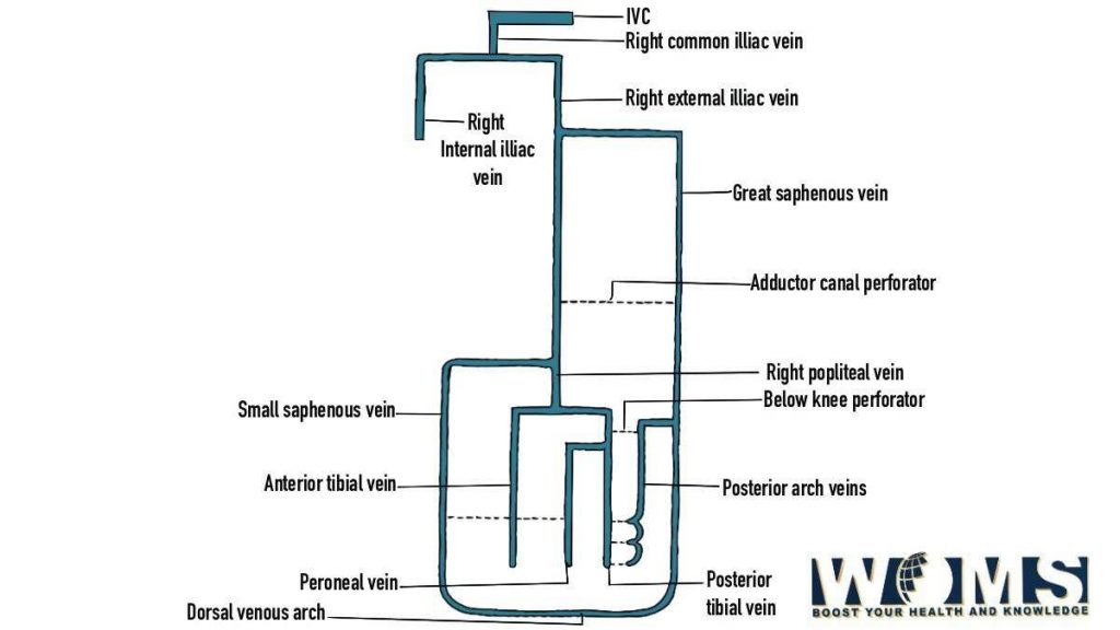
- Deep femoral vein – This blood vessels maintain its access at the rear of the femoral vein at about 8 centimeters from the inguinal ligament.
- Great saphenous vein – It is the body’s largest vein that extends from the feet to the thigh through the subcutaneous tissue. It unites with the femoral vein in the front region, closer to the pelvis area.
- Circumflex femoral veins – There are several medial and lateral circumflex femoral veins that also drain deoxygenated blood into the femoral vein
- Muscular tributaries – these tributaries also drain into the femoral vein.
Anatomical variants of the femoral vein
The femoral vein exhibits multiple structural variations due to different kinds of venous malformations. These variations in femoral vein anatomy develop during the late developmental phases of the embryo. These variations may be the number of femoral veins and the caliber of the main trunk of the vein in the thigh region. Here are some of the most common anatomical variations of the femoral vein anatomy with their prevalence.
- Axofemoral trunk – in this condition, the femoral vein fails to form properly. In this case, the axial vein is the major vein in the thigh region.
- Deep femoral trunk – This condition arises when the femoral vein fails to develop properly. In this condition, the deep femoral vein becomes the primary vein to drain blood from the lower limbs.
- Duplicated femoral vein (12 to 46%) – it is one of the most common variations in which a duplicate of the femoral vein also moves parallel to the original femoral vein.
Clinical importance of the femoral vein anatomy
Because of its increased diameter and width, the femoral vein offers great clinical significance during several surgical procedures. There are several clinical procedures that require proper care of the femoral vein. Let us take a rapid review of these clinical procedures and importance of femoral vein anatomy.
Venous Sampling
It is a very important tool to compile the sample of vein tissues for proper testing and assessment. This procedure helps the doctor to diagnose various hormonal problems like Cushing’s syndrome, hyperthyroidism, etc. This femoral vein is an important access point to collect the vein tissue to assess various diseases.
Deep vein thrombosis
It is a very common and potentially harmful disease related to the clotting of blood in the deep vein. This condition commonly occurs in veins of the lower limb like the femoral vein. It becomes harmful if the breakdown products of blood reach the lungs through blood, causing pulmonary embolism. This disease arises from various symptoms like swelling, tenderness, and pain. If not diagnosed earlier, it may cause increased heartbeat with breathing difficulty, light-headedness, and cough. It usually requires immediate surgical management.
Catheterization
During this surgical procedure, a small tube-like material is passed through the femoral vein to gain access to the right atrium of the heart. This process is done to diagnose various heart diseases like coronary artery disease, heart failure, etc. Femoral vein anatomy should be known well concerning these clinical importance.
The sound of blood flow in heart and lungs both are of high clinical significance. You can check Auscultation world app to hear clear and high quality heart and lung sounds.
Frequently asked questions (FAQs)
What are the common diagnostic tests to check the health of femoral veins?
Your doctor may offer you different diagnostic tests to confirm the health of the femoral vein. These are as follows:
● CT scan
● MRI
● Venous duplex ultrasound
What are the common symptoms associated with diseases of the femoral vein?
If there is any problem associated with your femoral vein, you may present to your doctor with the following symptoms.
● Pain in legs
● Itchy skin or other skin problems
● Swelling of the leg
● Varicose veins
● Ulcer formation