Ligaments of the Liver – Anatomy of Liver
Ligaments of the liver are the double-layered structures that are the foldings of the peritoneum. It surrounds the liver in such a way to connect the liver to the abdominal wall and different other body organs. Ligaments of the liver are usually the regressed particles of the embryological blood vessels that are resorbed as the embryo grows.
Ligaments of the liver are named according to the structures they connect with the liver. It helps the students to learn the anatomy of these ligaments quite rapidly. There are different ligamentous attachments of the liver that help in the anchorage of the liver on the right side of the upper quadrant of the peritoneal cavity. Moreover, these ligamentous attachments are as follows:
- Coronary ligament
- Triangular ligament
- Falciform ligament
- Ligamentum teres
- Ligamentum venosum
- Lesser omentum
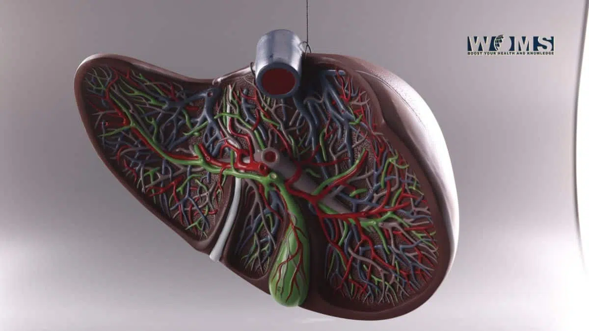
Anatomy of liver
The liver is the most important and second-largest organ of the human body. It is present below the diaphragm existing in the right upper and mid abdomen. Moreover, it also extends towards the left upper abdomen. The liver exhibits a typical prism or wedge shape, with its base lying to the right and apex towards the left. The superior surface of the liver can be percussed over the 5th intercostal space.
Anatomically, the liver is divided into the right larger lobe and the left smaller lobe. This division is demarcated by the presence of a falciform ligament.
Let us have a detailed look at the anatomical features of these ligaments of the liver.
Coronary ligament
The coronary ligament consists of anterior and posterior folds. As ligaments connect the liver to different other structures. It connects the superior and posterior parts of the liver’s right lobe to the lower aspect of the diaphragm. In addition, it is the largest ligament of the liver demarcating the diaphragmatic peritoneum. As we mentioned, it consists of anterior and posterior folds.
The anterior fold connects the superior aspect of the liver to the diaphragm towards the right-hand side.
The posterior fold connects the posterior aspect of the right lobe of the liver to the right side kidney, the abdominal wall on the posterior aspect, and associated suprarenal glands.
The coronary ligament is represented as the bare area of the liver. These anterior and posterior folds of the coronary ligament form a triangular area. This triangular region is without a peritoneum. Therefore, this region is also called the bare area of the liver. This ligament also plays a major role when it comes to ligaments of the liver under liver anatomy.
Triangular ligament
Triangular ligaments are bilateral structures present on the right and left sides of the liver. These right and left triangular ligaments are actually formed by the anterior and posterior layers of the coronary ligament. The main function of this ligament is to keep the liver in its place. It anchors the left lobe of the liver to the diaphragm. It starts from the apex point of the bare area of the liver.
The right triangular ligament is quite short. In contrast, the left triangular ligament is larger than the right. This ligament is present on the anterior side of the abdominal esophagus, cranial aspect of the lesser omentum, and medial part of the gastric fundus. The left triangular ligament extends along the liver for a larger extent. In this way, it becomes continuous with the lesser omentum and falciform ligament on its pathway. This is another ligament under ligaments of the liver.
Falciform ligament:
The falciform ligament is the second-largest ligament of the liver. It is a sickle-shaped ligament. It extends in a craniocaudal direction moving towards the anterior aspect of the liver. In the superior aspect, this ligament connects the visceral surface of the abdominal wall and the lower surface of the diaphragm. Majorly, it anchors the anterior aspect of the liver to the anterior wall of the abdomen.
Just like other ligaments of the liver, falciform ligaments also consist of two main layers. The right layer extends laterally to unite with the coronary ligament following the right side according to its name. Whereas, the left layer moves in a medial direction to merge in the triangular ligament on the left side. These two layers join to form a falciform ligament. It moves in a downward direction on the anterior aspect of the liver demarcating the division of lobes of the liver. In addition, this ligament also contains ligamentum teres, an embryological remnant of the umbilical vein. This is another important ligament under the ligaments of the liver
Ligamentum teres
Ligamentum teres is another ligament under the ligaments of the liver. It is the embryological remnant of the left umbilical vein that drains into the left portal vein. The left umbilical vein degenerates after about two months of birth. The remnants of this vein are known as ligamentum teres hepatis or ligamentum teres. This ligament usually connects the inferior and posterior surfaces of the liver to other organs of the body.
This ligament continues its pathway along the inferior aspect of the falciform ligament and extends its course along the fissure of the ligamentum venosum. Moreover, this ligament represents the inferior and posterior attachments of the liver.
Ligamentum Venosum
During the developmental phases, the ductus venosus was the main duct to provide a shunt pathway for the blood of the left portal vein towards the left hepatic vein. In this way, it helps to bypass hepatic circulation. In the first week of extrauterine life, this duct starts regressing. Ultimately, it forms the ligamentum. This is among the ligaments of the liver that is just like a fibrous cord that extends from the left portal vein in an upward direction and reaches the inferior vena cava. It is invested usually in the fissure of lesser omentum present on the visceral aspect of the liver.
Lesser omentum
The lesser omentum is among the ligaments of the liver that contains hepatoduodenal (liver to the upper duodenum) and hepatogastric (liver to the stomach) ligaments. Moreover, these ligaments of the liver connect the liver to the upper part of the duodenum and the lower aspect of the stomach. It is an “L” shaped ligament extending from the distal part of the esophagus moving along the inferior curvature of the stomach and ultimately connecting the upper portion of the duodenum.
The Hepatogastric ligament tracks the distance between the stomach and liver. It contains two main layers, merging with the left triangular and right coronary ligament. It moves forward to encroach on the inferior vena cava.
The hepatoduodenal ligament connects the liver and the upper part of the duodenum. It surrounds the main structure of porta hepatis including the portal vein, hepatic artery, and bile duct.
Clinical importance of the ligaments of the liver
These ligaments of the liver encode great clinical considerations prior to any medical procedure. There are several clinical aspects that remind the importance of these ligaments. Besides these, you should also know about Portal trial and essential structures passing it to know the clinical interpretation better.
Let us look at these clinically important points to understand.
- In surgical procedures that need the mobilization of the liver, triangular and right coronary ligaments are usually separated to freed the right lobe of the liver. It is also important to achieve access to the region of the retrohepatic area up to the inferior vena cava.
- In right hepatic lobectomy, the left triangular ligament is important to consider clinically. If any surgeon is unable to repair this ligament after hepatic lobectomy, there will be a prominent imbalance in the liver’s left lobe. It may also lead to kinking of liver veins with liver dysfunctioning.
- In a pneumoperitoneum condition, a significant amount of air becomes entrapped in the abdominal cavity. Moreover, you can also observe a falciform ligament with free air outlining it on radiographic imaging. It indicates the presence of pneumoperitoneum.
- In a few cases, there is a failure in the degeneration of ligamentum teres hepatis. The patency of this condition is exacerbated resulting in Portal hypertension.
- Ligamentum Venosum is a primary landmark and a controlling source for the hepatic vein during surgeries that require proper control in the vessels.
Frequently Asked Questions (FAQs)
What is the main function of these ligaments of the liver?
These ligaments work synergistically to connect the liver lobes to the other structures and organs of the abdominal cavity. In addition, these ligaments are present in different areas of the liver to provide support from different regions.
Which ligament of the liver separates the lobes of the liver?
The falciform ligament is the ligament that is present between the lobes of the lobes. It helps to differentiate the lobes of the liver either right or left.