8 Cranial bones: anatomy, functions, and important clinical conditions
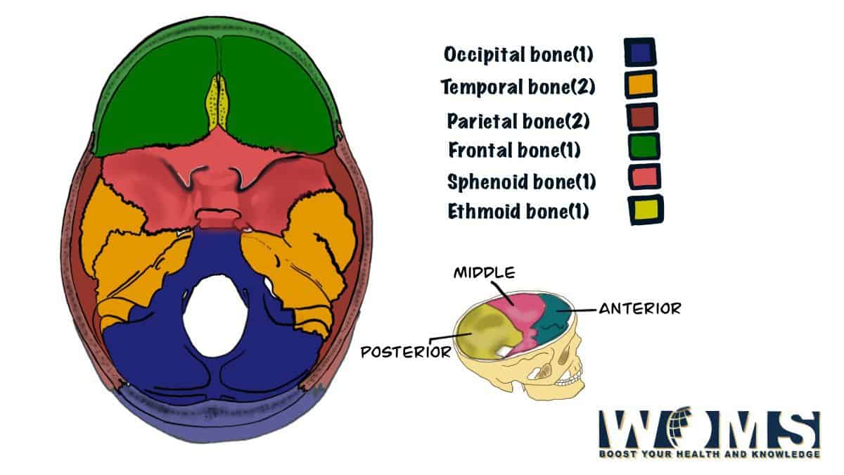
There are 8 Cranial Bones that form the enclosure of the brain. The frontal bone, two parietal bones, two temporal bones, the occipital bone, and ethmoid and sphenoid bones. The thickness of these bones varies and mainly depends on their position relative to the pterygopalatine fossa (sinus cavity in the back of the nose). The average thickness of the bones is around 3 mm.
The skull consists of many bones grouped into collections called axial (the connection between the brain and spine) and facial (subdivisions for nose, palate, cheekbones, and eye sockets), which make up a whole. We will focus on the important 8 cranial bones. The frontal bone is also axial as it connects to the forehead, a frontal sinuses cavity that communicates with your nose. The shapes of these surfaces can be used to identify age and gender.
8 Cranial Bones
Let us discuss each of the 8 cranial bones in detail. All the 8 cranial bones are discussed briefly below:
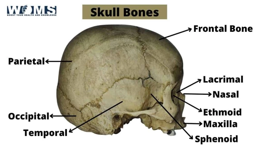
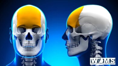
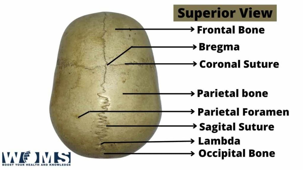
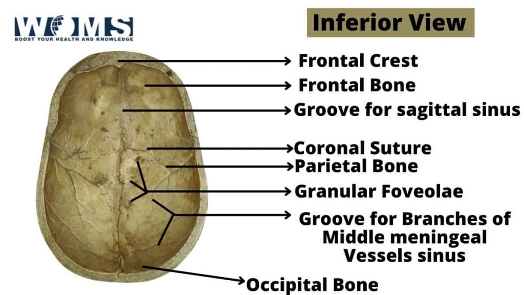
1. Frontal bone
The frontal bone is the anterior-most bone in the occipital bone, forming an elongated structure that separates the frontal sinus from the middle cranial fossa. The frontonasal sinus (a small cavity found just below your nose) communicates with this space by way of a small tunnel which widens at its base.
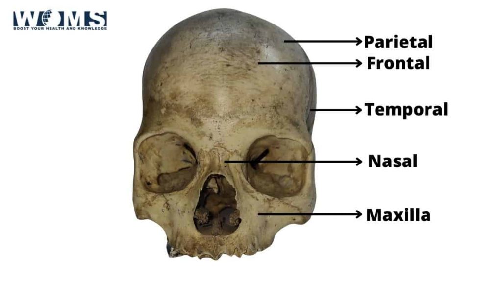
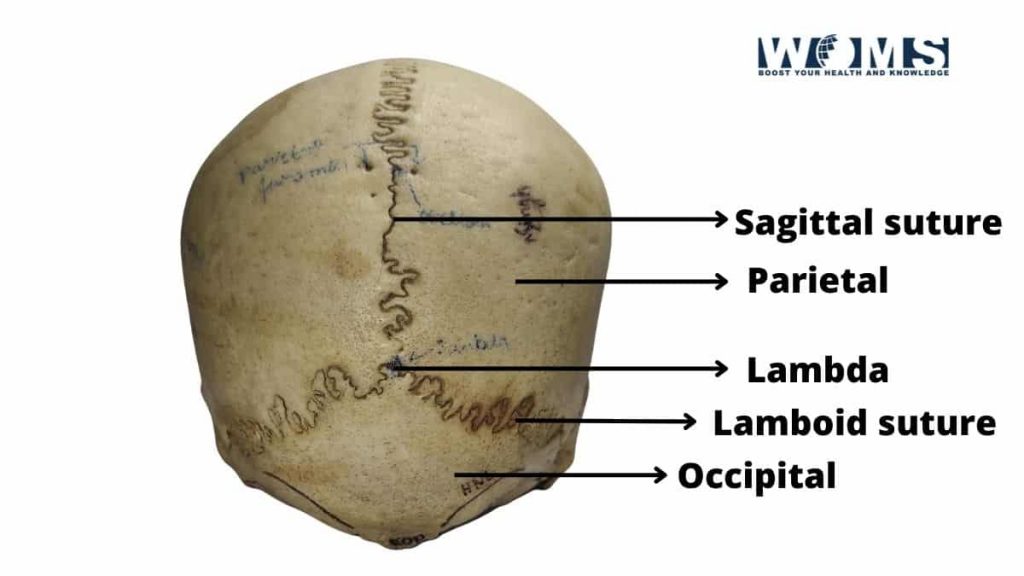
2. Parietal bones (Paired)
The parietal bones form part of the side of your head (including eye sockets). These bones help to support your face and keep it straight. The brain is located between the parietal bones on the floor of the middle cranial fossa. The top portion of the parietal bones forms your frontal and parietal opercula, which contain regions responsible for hearing and balance.
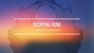
3. Occipital bone
The occipital bone is located posterior to the parietal bones and has two plates that support our skull’s base. The two occipital plates form a bony arch over our ears that allows for more flexible movement and improves hearing. The underside of this bone forms part of your ear, giving us better hearing (and a giant ear opening for sound waves to enter).
4. Temporal bones (Paired)
The temporal bones are located behind the parietal bones, and the zygomatic bone forms its temporomandibular joint (jaw bone). The temporal bones form the sides and middle of your head, including the temples, ear holes, and parts of your face. The temporal bone also contains many vital structures, such as the auditory nerve (facial nerve), which helps send sound from your tympanic membrane to your brain.
5. Sphenoid bone
The sphenoid bone is on the same side of your face as the ethmoid bone. It is a ball-like central part of the upper jaw and forms part of many facial features, including our cheekbones and nose.
6. Ethmoid bone
The ethmoid bone is the outermost roof of the nose and forms vital olfactory nerves that detect the smell. This bone also helps to create the orbit of our eye and other facial features. The ethmoid also contains three small cavities: the frontal, sphenoidal, and maxillary sinuses. Abscesses can form in these cavities when they become infected.
Key Facts
Landmarks: Temporalis, masseter, anterior tubercle of occiput.
The Spinous process (C1) and mandibular condyle (C0) are other landmarks.
Cranial Nerves
All the 8 cranial bones get their nerve supply. The most commonly damaged cranial nerve is the fifth. This is because this part of the brain connects to the larynx at the top of your mouth and can cause damage if it is hit directly. Damage to this nerve will result in a temporary loss of ability to breathe.
Muscles
Besides the 8 cranial bones, the muscles in this region are discussed below:
1. Rhomboideus muscle
The Rhomboideus muscles are named after the Greek letter “rho” (ρ), a tiny jumping midline trapezoid-shaped muscle in the back of your neck.
2. Scalenus anterior
The scalenus anterior muscles are named for their superficial location just below the mandibular angle of your jaw and above your first rib. These muscles form part of the jaw and give it strength.
3. Scalenus medius
The scalenus medius muscles are located between the anterior and posterior scalenus. These muscles are shaped like a fan and run from your thoracic vertebrae to your first rib.
4. Scalenus posterior
The scalenus posterior muscles are the deepest of the scalene muscles, and they form the front side of your neck. These muscles also run from your thoracic vertebrae to your first rib.
5. Rectus capitis anterior
The rectus capitis anterior muscles are located on the neck’s upper part and help bring your head back.
6. Rectus capitis lateralis
The rectus capitis lateralis muscles are located on the lower portion of your neck and help to turn your head to the side.
7. Rectus Capitis posterior major
The rectus capitis posterior major is located at the base of your skull by your occipital bone. These muscles help to bring your head back and are responsible for rotation.
Joint
The information about the 8 cranial bones won’t be enough without knowing about the joints. Here are the joints that you need to know about:
1. Craniovertebral joint
The craniovertebral joints or craniovertebral joints are located where your brain and spinal cord attach to the top of your spine. This joint helps to provide flexibility in your head, allowing it to be able to bend and twist.
2. Temporo-mandibular joint
The temporomandibular joint is located where the mandible (lower jaw) and the temporal bone form a disc. This joint helps to develop the lower jaw and allows for movement.
3. Zygomatic joint
The zygomatic joint is where the zygomatic bone forms part of your cheekbone, and the temporal bone joins. This joint allows for movement of your jaw, cheeks, and nose.
4. Maxillary joint
The maxillary joint is where the maxilla (upper jaw) and the zygomatic bone form a disc. This joint allows for movement of your lips, cheeks, and nose.
5. Mandibular joint:
The mandibular joint is located where the mandible (lower jaw) and the maxilla (upper or lower jaw) form a disc. This joint allows for your lower jaw, lips, and cheeks movement.
These are some important joints between the cranial bones including the important 8 cranial bones that we discussed.
Clinical Relation
These 8 Cranial bones may be related to clinical conditions such as migraine and lower-limb paresis. Cranial bones can be fractured due to traumatic injury. Clues to the possibility of a fracture include: rapid loss of consciousness, nausea, vomiting, headache, diplopia (double vision), drowsiness/lethargy/sluggishness, loss of balance and falling, decreased motor function, or paralysis. If a fracture has occurred, there is an abnormal sensation over the affected site that may include pain or tenderness.
Symptoms
If the 8 cranial bones that we discussed undergo fracture, the patient experiences some symptoms. The patient will experience extreme pain and swelling of their arm, neck, and face. The fractures are often painful to the touch. As the jawbone heals during healing, the patient will notice stiffness or a lack of motion in their jaw.
Causes
Children are more prone to break the jaw or teeth due to the enlargement of the growth plates by 2-3 times compared to adults. The jawbone is, therefore, more susceptible to fractures.
This was the basic clinical correlation including that of the 8 cranial bones.
Conlculsion
All the 8 Cranial bones are unique because they are the only skull bones not joined together by immovable joints. They move independently from each other and provide a wide range of motion. This allows for multiple functions such as chewing, breathing, swallowing, balance, and the ability to speak, among many others. The cranial bones allow for movement of our facial features and jaw, forming perfectly with the rest of our skull to protect our brain from injury.
1. Why do children have different growth rates regarding their jaw bones?
The jawbone is an essential part of our face. The jaw is made of a set of small bones that are connected. One of the most noticeable differences between a child’s and an adult’s jaw is that the child’s growth plates are much larger and can grow for 2-3 times longer before closing than an adult’s. The growing ends tend to be much wider than in adults, making them more susceptible to injuries from injury.
2. Discuss the difference between “growth plate” and “growth cartilage” and recommend materials you would use to help manage fractures.
Growth plates are tiny spongy areas at the end of a bone that negatively affect bone growth. The growth plates can be soft, but they are essential to bone development as they are where new bone is grown. The bones in the face and skull begin their growth in these areas, which is why they are often more sensitive to injury and fracture than other areas. Growth cartilage is a softer area found between the bones at the extremities (e.g., elbows, knees, etc.).
Why do children suffer some common fractures?
One of the most common fractures a child suffers involves their growth plates. Growth plates are areas of soft tissue that can not only be damaged but are often the most sensitive areas on a bone. They cause a lot of swelling as they heal, making them more susceptible to breakage.