Vertebral Foramen : structures, functions, and clinical significance
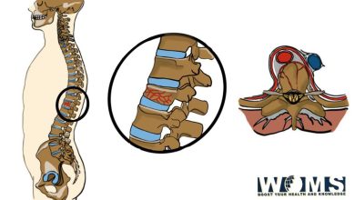
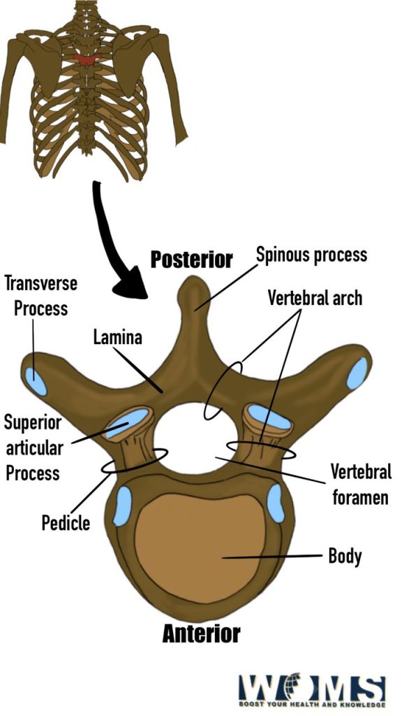
The vertebral foramen is a potential space between the spinal cord and the posterior elements of the spine, through which nerves pass from the spinal cord to the rest of the body. The vertebral foramen is located below the neural arch (the part of the vertebra that contains the spinal canal) and above the transverse process. It can be divided into three parts: anterior, middle, and posterior. Each has a different function.
The anterior portion of the vertebral foramen is formed by the laminae and spinous processes of the vertebrae. The pedicles and transverse processes form the middle portion. The articular facets form the posterior portion. The nerve roots enter the vertebral foramen through the posterior portion. The vertebral artery passes through the vertebral foramen in the middle portion.
The vertebral foramina anatomy consists of two lateral recesses and one medial recess. Two openings are also on each side of the foramen, called intervertebral foramina. These openings allow passage of the spinal nerves and vessels. They are located at the junction of the superior and inferior articular processes. The lateral recesses are larger than the medial recess. The lateral recesses contain the exiting nerve root fibers. The medial recess contains the ascending branch of the dorsal ramus of the spinal nerve.
The function of the vertebral foramen
The vertebral bodies are hollow bony cylinders containing cancellous bone tissue enclosed within tough outer layers of dense cortical bone. The vertebral bodies have a central cavity, known as the vertebral body’s “canal.” This cavity extends from the center of the vertebral body to its periphery. The vertebral body’s posterior wall is reinforced with strong bands of fibrocartilage known as annulus fibrosis. The annulus surrounds the vertebral body and encloses the spinal cord and the cauda equina. The annulus is composed of concentric rings of collagenous fibers and elastic lamellae.
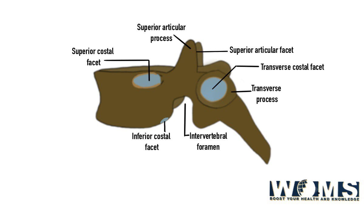
Structures of vertebral foramen
Four types of bones form the vertebral column: the vertebral bodies, the intervertebral discs, the facet joints, and the ligaments. The vertebral bodies are cylindrical bones that house the spinal cord and the nerve roots. The intervertebral discs separate the vertebral bodies and act like shock absorbers. The facet joints connect the vertebral bodies and allow them to move relative to each other. Finally, the ligaments hold the vertebral bodies together.
The vertebral bodies consist of cancellous bone surrounded by a thin shell of compact bone. The cancellous bone is richly vascularized and contains many small air cells. The compact bone of the vertebral body is denser than the surrounding cancellous bone. Its structure allows it to withstand compressive forces without deforming.
The intervertebral disc
It is a soft, gelatinous cushion that lies between the vertebral bodies. The intervertebral is comprised of three distinct regions: the nucleus pulposus, the annulus fibrosus, and the cartilaginous end plates. The annulus fibrosus is a thick, multi-layered band of dense fibrous tissue that encircles the nucleus pulposus. It acts much like a laminated plate in a door hinge. The annulus fibrosis prevents the nucleus pulposus from protruding into the spinal canal.
Nucleus Pulposus
The nucleus pulposus is a gel-like substance within the disc’s center. It has no cellular components and consists mostly of water, some proteoglycans, glycoproteins, and hyaluronic acid. The nucleus pulposus provides most of the mechanical support for the disc. It also serves as a reservoir for nutrients transported to the disc by way of the blood vessels in the endplate.
Annulus Fibrosus
The annulus fibrosus comprises an outer ring of strong, flexible fibers surrounding the softer nucleus pulposus. As mentioned above, this ring functions like a laminated plate door hinge. The annular fibers attach to the endplates of adjoining vertebrae and provide stability to the motion segment.
Cartilaginous End Plates
The cartilaginous end plate covers the surface of the opposing vertebral bodies and attaches to the inferior articular processes. These end plates contain numerous pores through which synovial fluid can flow into the joint space.
Facet Joints
These joints are located at the junction of the lamina and transverse process of one vertebra and the spinous process of the next. They are covered by smooth, hyaline cartilage. The facets prevent the vertebral bodies from moving too far apart or coming closer together. Facet joints are not directly connected to the rest of the spine but have attachments to the posterior elements.
Ligaments
These structures serve two main purposes: (1) they stabilize the vertebral bodies, and (2) they limit the movement of the vertebral bodies. Ligamentum flavum: This ligament connects the upper part of the vertebral arch to the spinous process. It helps keep the cervical spine stable and protects the spinal cord. Interlaminar Segments: These segments of the spine lie between adjacent pairs of vertebrae. They help maintain the normal distance between the vertebral bodies and protect them from injury.
Nerves of the vertebral foramen
Many nerves pass through the vertebral foramen. These include the anterior primary rami of C5 and T1, the lateral primary rami of C6 and T2, and the medial primary rami of C7 and T3.
Treatment of Spinal Disorders
In general, treatment of spinal disorders involves reducing pain, restoring mobility, and preventing further damage to the patient’s nervous system. Treatment may involve medication, physical therapy, injection therapies, and surgery. Medication is typically used first to treat acute symptoms and reduce inflammation. Physical therapy is often prescribed after the initial recovery period. Surgery may be required if conservative treatments fail.
Joint of vertebral foramen
A joint of the vertebral foramen is formed when the superior articular facet of one vertebra articulates with the inferior articular facet of another vertebra. A joint of the vertebral foramen is a type of synovial joint. Synovial joints allow relative movement between bones while limiting the amount of translation and rotation possible between those bones. In other words, synovial joints permit some degree of freedom of movement within certain limits.
Synovial joints are classified as either diarthrodial or amphiarthroidal. Amphiarthroidal joints are also known as “ball-and-socket” joints because they have a ball-shaped bone in one member (the socket) and a corresponding spherical indentation in the other member (the ball). Diarthroidal joints are characterized by a saddle shape where each bone has an indentation that fits over the top of a ridge on its counterpart.
Amphiarthroidal joints are found in the wrist, elbow, knee, ankle, and foot. Examples of amphiarthroidal joints include the hip, shoulder, and intervertebral disc.
Diarthrodial joints are called hinge joints because the joint surfaces move around a fixed axis. Hinge joints are found in the fingers, toes, and the temporomandibular joint (TMJ).
The TMJ is a complex joint consisting of three bones: the mandible (lower jaw), the temporal bone (upper jaw), and the glenoid fossa of the temporal bone. The TMJ has four major joints: condylar, central, capsular, and disk. The condylar joints are located at the front of the head and neck. Each condyle is covered by cartilage and surrounded by fibrous tissue. The major joints connect the two halves of the skull.
Clinical Significance of vertebral foramen
The vertebral foramen is a space that lies between the posterior aspect of the lamina and the neural canal. It contains the nerve roots and blood vessels that supply the spinal cord. The vertebral foramen provides a pathway for transmitting mechanical forces and signals along the spinal column.
Spinal Canal
The spinal canal is a skinny tube that extends from the base of the skull through the spine to the sacrum. The spinal canal houses and protects the spinal cord and nerves. The spinal canal allows the passage of nutrients and waste products into and out of the body via the spinal venous plexus.
Conclusion
The vertebral foramen is a very important part of our body. It is not only a passageway but also plays a vital role in protecting the spinal cord and nerves from injury.
Frequently Asked Question on Vertebral Foramen
How does the vertebral foramen protect the spinal cord and nerves?
• The vertebral foramina provide a pathway for the transmission and distribution of mechanical forces and signals throughout the spinal column.
• The vertebral foramina contain the spinal nerve roots and blood vessels that nourish the spinal cord.
• The vertebral foramen provides a pathway for transferring nutrients and waste products to and from the spinal cord.
• The vertebral foramen provides a pathway to transfer and distribute electrical impulses along the spinal column. This pathway is important for the control of muscle movement.
Why do you need to know about the anatomy of the vertebral foramen?
Knowing the anatomy of the vertebrae helps us understand the spinal cord’s position and its relationship with other structures such as the dura mater and ligaments.
What is the clinical significance of the vertebrate foramen?
Knowledge of the vertebral foramen anatomy may help understand the pathophysiology of various disorders affecting the spinal cord or the peripheral nervous system.
When should we consider vertebrate foramen when treating patients?
An orthopedic surgeon specializing in spinal surgery should evaluate a patient with symptoms of compression of the spinal cord or the cauda equina.
What are some common diseases involving vertebrate foramen and their effects on the patient?
Diseases involving the vertebral foramen include tumors (such as meningioma), infections (such as tuberculosis), and trauma (such as fractures). These conditions cause compression of the spinal cord and the cauda equina, resulting in neurological deficits.