Uncovering Complexities of the Horse Skull: An Anatomical Journey
Horses belong to the Equidae family, a taxonomic family including horses, zebras, and asses. In addition, there are several other extinct species belonging to this Equidae family. The anatomical details of the equine family are relatively similar. This article explores the gross and histological (microscopic) aspects of the horse skull and also other equids. Let us have a detailed review of the critical anatomical components of the horse skull to understand the clinical aspects related to the horse skull.
Which type of skeleton do horses have?
Horses contain two types of skeletons. These are
- Axial skeleton
- Appendicular skeleton
Axial skeleton
The axial skeleton of the horse contains a skull, vertebral column, sternum, and ribs. However, the skull covers the brain and also protects several other essential structures of the cranial cavity. We will discuss the horse skull in detail in the following sections. The vertebral column of the horse contains almost 54 bones to maintain the structural integrity of the horse. These 54 bones are in the form of vertebrae.
- Cervical vertebrae are 7 in number, including atlas C1 and axis C2.
- There are 18 to 19 thoracic vertebrae surrounding the region of the thoracic cavity.
- There are 5 to 6 lumbar vertebrae.
- Moreover, there are 5 sacral vertebrae that join to form a complete sacrum bone.
- There are 15 to 25 caudal vertebrae, with an average of 18 caudal vertebrae.
The differences in the number of these vertebrae mainly depend on the breed.
The sternum contains several sternebrae that fuse to form a single sternum bone. There are a total of 18 pairs of ribs. Moreover, the sternum is connected to 8 true pairs of ribs.
Appendicular skeleton
The appendicular skeleton holds the forelimb and hindlimb. The forelimb is not directly attached to the spine. It is in a suspended form attached by tendons and muscles. This structural arrangement allows a greater movement of the forelimb. The hindlimb of the horse is connected to the vertebral column through the pelvis. Hindlimbs maintain about 40% of the body’s weight. It is stabilized through various muscular attachments of the spine.
What are the important structures of the horse skull?
The horse skull contains the brain in a strong cranium. The skull protects the brain due to its bony structure.
Cranium or skull
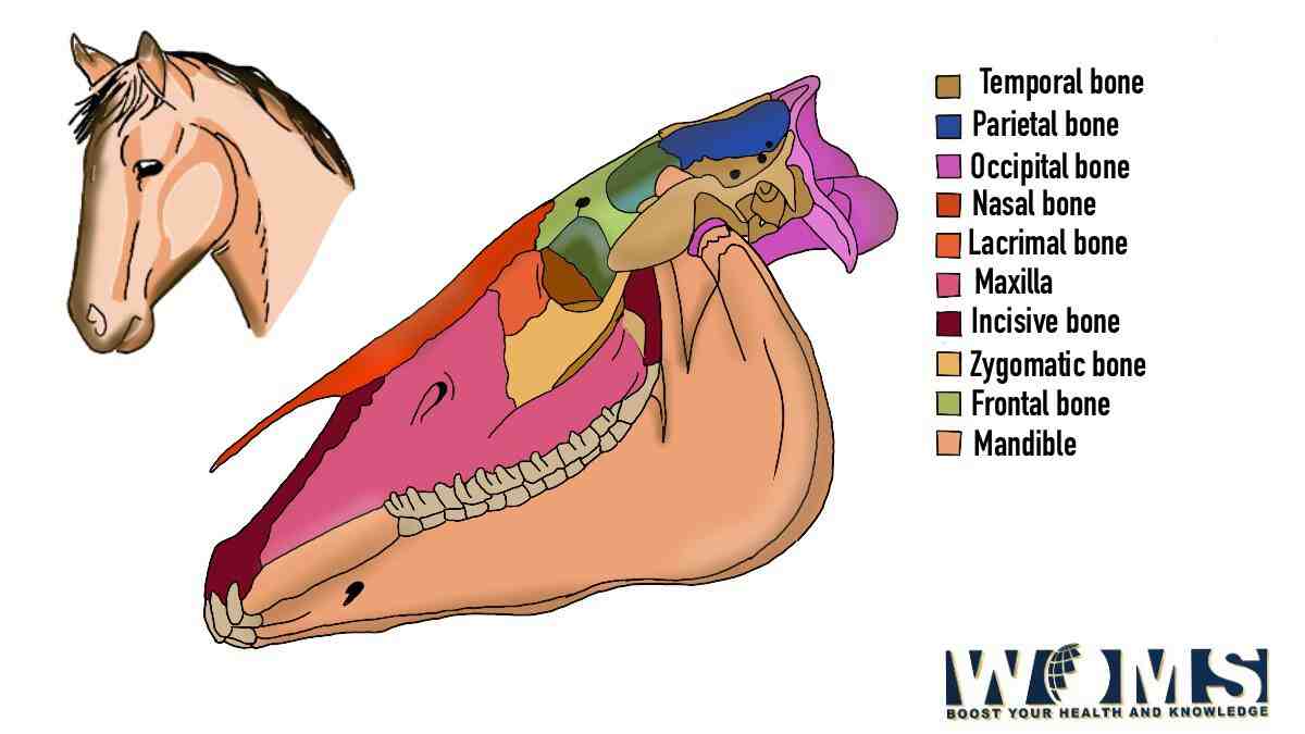
Cranium is made up of different bones, joining together to form a complete horse skull. The roof of the skull contains frontal and parietal bones. In addition, the floor of the cranium is made up of sphenoid bone. There are 34 bones in total that form the horse skull. These bones are flat and overlap with each other during the birth process. It makes the birth process easy by compressing the skull as much as possible.
The horse skull contains 14 major bones. These are as follows:
- Incisive bone – this bone is the anterior part of the upper jaw. It is the bone where the maxillary anterior attach.
- Nasal bone – as the name indicates, this bone surrounds the nasal cavity and offers a proper respiratory inlet to inhale and exhale.
- Maxillary bone – it is the larger bone that provides a proper facial orientation. In addition, it also surrounds the roots of upper molar teeth.
- Mandible – it is the largest bone of the horse skull forming the lower jaw. In addition, it also offers a proper bony structure to hold the roots of lower teeth in position.
- Lacrimal bone – this bone holds the nasolacrimal duct to provide a proper passage for fluids. It carries fluids from the nose and different surfaces of the eye to pass from.
- Frontal bone – this bone forms the forehead of the horse skull.
- Parietal bone – this bone extends from the forehead region of the horse skull and moves along the back of the skull.
- Occipital bone – this bony structure forms a joint-like connection between the skull and atlas, the first vertebrae of the neck.
- Temporal bone – this bone surrounds the external acoustic meatus to transfer the sound from the ear to the eardrum or cochlea.
- Zygomatic bone – this bone creates the prominent part of the cheekbone. In addition, it is attached to the temporal bone.
- Palatine bone – it is a palate bone that forms the posterior part of the hard palate.
- Sphenoid bone – this bone is formed by the combination of the fetal basisphenoid and presphenoid bones. It forms the base of the skull. It can be fractured in those horses that rear over backward.
- Vomer bone – this bone forms the upper part of the interior surface of the nasal cavity.
- Pterygoid bone – it is a small bone connected to the sphenoid bones that move downward.
What are the important cavities of the horse skull?
The horse skull consists of four important cavities. These are as follows:
Cranial cavity
It surrounds and provides a protective barrier to the brain. As the brain contains a variety of sensory stimulation, the cranial cavity protects all the important sensory organs. The roof of the cranial cavity is formed by the parietal and frontal bones. Whereas the floor of the cranial cavity is made up of sphenoid bone.
Orbital cavity
The orbital cavity surrounds almost five orbits formed by frontal, lacrimal, sphenoid, palatine, and zygomatic bones. These bones protect and cover the eye‘s contents. Horses possess both monocular and binocular types of vision.
- Monocular vision – means that a horse can see different objects with one eye. In this type of vision, the horse’s brain receives two images simultaneously.
- Binocular vision – it is quite similar to human eyesight. Horses can focus on objects with both eyes, similar to humans. But, they receive only one signal.
Oral cavity
The oral cavity is a way to both the respiratory and digestive systems. It is also formed by different bones to protect the inner organs of the body.
Nasal cavity
The nasal bone surrounds the nasal cavity. It protects the mucous membranes that maintain the optimum temperature of inhaled air.
Also, read: Understanding Dog Ear Infection Medicine
What are the important foramina of the horse skull and associated structures passing through them?
The horse skull possesses multiple foramina to pass different structures from the inside of the skull to the outside. These foramina are the linking structures that connect the inner response to the outer stimulus. These foramina and their associated structures are as follows:
- Infraorbital foramen – passage for infraorbital nerve CN-V
- Maxillary foramen – it also makes a way for cranial nerve V (infraorbital nerve).
- Cribriform foramen – it offers a way for the olfactory nerve which is cranial nerve I.
- Optic canal – this canal provides a passage for the optic nerve which is cranial nerve II.
- Orbital fissure – this fissure offers a passage for different cranial nerves to pass. The cranial nerve IV, V, VI (ophthalmic division), and VII passes through this fissure.
- Round foramen – this foramen makes a way for CNV (maxillary division).
- Oval foramen – this foramen delivers cranial nerve V (mandibular division).
- Foramen lacerum – this foramen makes a way for the internal carotid artery and cranial nerve V3.
- Internal acoustic meatus – this opening passes cranial nerve VIII outside the skull.
- Jugular foramen – this foramen helps to move cranial nerves IX, X, and XI to the outside of the skull.
- Stylomastoid foramen – this foramen makes a passage for cranial nerve VII.
- Mandibular and mental foramen – these foramina are the passing ways for cranial nerve V ( mandibular alveolar nerve).
What are the important joints and ligaments associated with a horse skull?
There are different joints and ligaments associated with the horse skull. Joints allow a slight movement of the bones. These joints are as follows:
- Temporomandibular joint – as the name indicates, this joint is present between the mandibular condyles and the associated mandibular fossae of the temporal bone. It is a condylar joint with a loose joint capsule and thickenings to form a lateral ligament. In addition, it also contains an articular disc.
- Mandibular symphysis – it is located near the rostral end of the mandible connecting the right and left halves of the mandible.
- Hyoid apparatus – hyoid apparatus is formed by three joints which are as follows:
- Between the tympano-hyoid cartilage and skull
- Interhyoid joints
- Between the thyrohyoid bone and cranial cornu of the thyroid cartilage
The horse skull contains a variety of muscles to orient different functions. There are specific muscles associated with the specific structure to deliver specific responses through these muscles. Let us have a look at these muscles and their functions accordingly.
Muscles of the face
- M. Levator labii maxillaris – it helps in the elevation of maxillary lips.
- M. Levator nasolabialis and M. Zygomaticus – these muscles help in the elevation and retraction of the angle of the mouth.
- M. Buccinator – this muscle helps in the flattening of the cheeks and provides a pressing force for the food between the teeth.
- M. Depressor labii mandibularis – it depresses and retracts the mandibular lip.
- M. Risorius – it also helps in the retraction of the angle of the mouth.
- M. Dilator naris – it dilates the nostrils.
- M. Lateralis nasi – it helps to dilate the nostrils and nasal vestibule
- M. Caninus – it helps in the dilation of the nostrils laterally.
- M. Levator nasolabialis – it elevates the maxillary lip and commissure or the mouth. Moreover, it also helps to dilate the nostrils.
Muscles of mastication
- M. Masseter – it helps to close the mouth.
- M. Temporalis and M. pterygoideus medialis – these muscles help to close the mouth by raising the mandible in an upward direction.
- M. Pterygoideus lateralis – this muscle moves and dra3e the mandible in the rostral direction.
- M. Digastrucus and M. Occipitomandibularis – these muscles help to open the mouth.
Muscles of the eyes
- M. Orbicularis Oris
- M. Levator palpebrae superioris – elevates upper eyelid
- M. Malaris – lowers the ventral eyelid
- Muller’s muscle
- Ciliary muscles
- M. Retractor anguli – anchors and retracts lateral canthus
- M. Levator anguli oculi medialis and M. Frontalis – slightly elevates the upper eyelid
Muscles of the tongue
The horse tongue is made up of 12 different kinds of muscles including styloglossus, hyoglossus, geniohyoideus, and genioglossus. These muscles are fully covered by the mucosa.
Conclusion
The horse skull consists of a combination of different bones. Moreover, these bones are joined together with the help of joints. In addition, there are several foramina to maintain a link between the contents inside the skull and outside of the skull. Moreover, the brain inside the horse skull is a hub for a variety of sensations in the body. The skull protects the brain and indirectly the complete metabolism of the horse body. There are also several muscles associated with the horse skull that help to perform multiple functions.
This article described complete details related to the horse skull and associated structures, bones, joints, and muscles. Go through the article in detail to clear your anatomical concepts related to the horse skull.
Frequently asked questions (FAQs)
What is the unique factor about the horse skull?
The unique factor related to the horse skull is the number of bones making up the horse skull. The horse skull is made up of 34 different bones in comparison to the human skull. The human skull is made up of 14 bones. Therefore, the horse skull is different in aspects of size and shape.
Which is the largest bone in the horse skull?
Comparing the bones of the horse skull, the mandible is the largest bone in the horse skull.