Cases
Supracondylar Fracture of Humerus
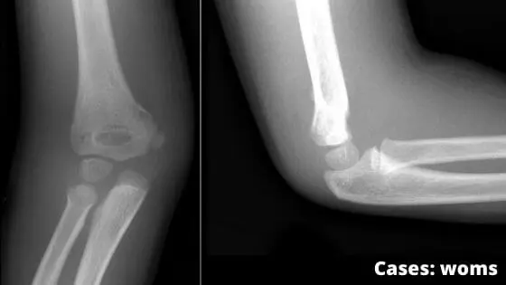
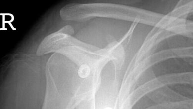
A 55-year old individual sustained a severe blow on his right flexed elbow. He developed pain and swelling in the elbow region. He was taken to an orthopedic surgeon who on examination found that the three bony points (olecranon, medial epicondyle, and lateral epicondyle) in the below region were forming an equilateral triangle. He suspected a fracture and advised the x-ray of the elbow region. The x-ray revealed the supra-epicondylar fracture of the humerus.
Questions
- What are the three common sites of the fracture of the shaft of the humerus? Name the nerves related to these sites?
- Why the triangular relation of three bony points (olecranon, medial epicondyle, and lateral epicondyle) is not disturbed in the supracondylar fracture of the humerus.
- Which is the most commonly injured nerve in the supracondylar fracture of the humerus?
- On clinical examination, how will you differentiate the supracondylar fracture of the humerus from the posterior dislocation of the elbow?
Answers
- Common fracture sites of the humerus:
- a) Surgical neck – associated with injury to the axillary nerve
- b) Mid-shaft (radial groove region) – associated with injury to the radial nerve
- c) Supracondylar region – most commonly associated with injury to the median nerve (especially the anterior interosseous branch)
- In the fixed elbow, the three bony points of the elbow (olecranon, medial, and lateral epicondyles) from an equilateral triangle/ isosceles triangle. The triangular relationship remains intact in a supracondylar fracture because the fracture occurs proximal to the medial epicondyle, lateral epicondyle, and olecranon. Therefore, the anatomical relationship among these bony landmarks is preserved.
- The median nerve is the nerve most commonly injured in supracondylar fractures. The anterior interosseous branch may be particularly vulnerable.
- In a supracondylar fracture, the triangular relationship between the olecranon and the medial and lateral epicondyles remains normal. In posterior dislocation of the elbow, the olecranon is displaced posterosuperiorly, resulting in loss of the normal triangular relationship.
Key point: Along with checking bony landmarks, always assess motor and sensory function of the median, radial, and ulnar nerves. This helps identify any associated neurovascular injuries early.