Cases
Median Cubital Vein
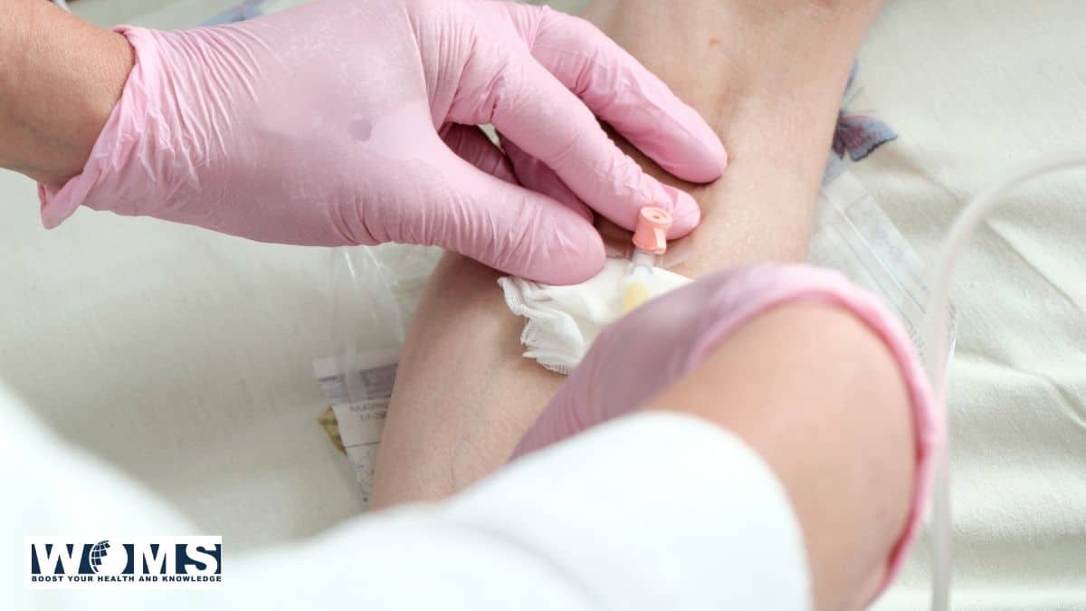
A 38-year-old female visited a clinic for a routine blood examination. The pathologist asked the technician to collect the blood sample of the lady. While attempting to collect the blood sample from the median cubital vein the technician noticed that the blood in the syringe is bright red. He immediately withdraws the needle.
In the second attempt, he inserted the needle slightly medial to the previous puncture. The lady felt a sharp pain, which radiated to the lateral three digits.
Questions:
- What is the median cubital vein?
- Name the fascial structure, which separates the median cubital vein from brachial artery and median nerve.
- Mention the cause of the sharp pain that radiated to the lateral three and a half digits.
- What does the bright red blood in syringe indicated during collection of the blood sample from the median cubital vein?
Answers:
- It is a communicating vein in front of the elbow between the cephalic and basilic vein.
- Bicipital aponeurosis. (The bicipital aponeurosis runs obliquely downwards and medially from the biceps tendon, blending with the deep fascia of the forearm. It protects the underlying brachial artery and median nerve from accidental punctures during routine venipuncture, acting as a crucial anatomical shield.)
- Median nerve injury. (The median nerve lies immediately medial to the brachial artery in the cubital fossa; moving the needle medially caused direct trauma to this nerve.)
- Accidental puncture of the brachial artery. (Bright red, pulsating blood indicates highly oxygenated arterial blood, rather than the dark red deoxygenated blood typically found in the median cubital vein.)