Scapula: Anatomy and Clinical Relevance
The scapula is also called the “shoulder bone”. (Scapula is a Latin word that means ‘shoulder blade’). Let’s learn the scapular anatomy in detail.
The scapula is a wide and flat bone in the shape of a triangle that rests on the posterior thoracic wall. It is located between the second and seventh rib. It connects with the acromial end of the clavicle and the head of the humerus laterally.
The body of the scapula bone is thin although its borders, especially the lateral border, are thicker.
The major defining characteristics of this bone are its:
- Three borders (superior, medial, lateral)
- Three angles (Superior, inferior, lateral)
- Two surfaces (dorsal, coastal)
- Three bony prominences (scapular spine, acromian, coracoid)
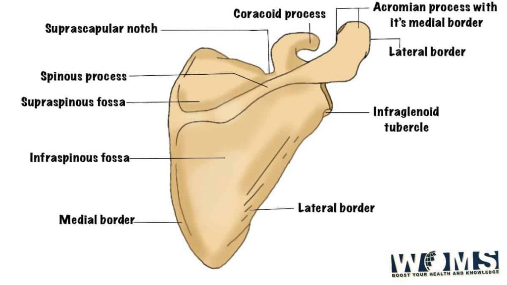
Superior Border of Scapula
The superior border is a short and fine margin of the scapula. It is located at the junction of its medial two-thirds and the suprascapular notch laterally.
Suprascapular notch
It is located at the junction of the superior border and coracoid process. The superior transverse ligament passes over the notch and the suprascapular artery traverses superior to this ligament while the suprascapular nerve lies inferior to it.
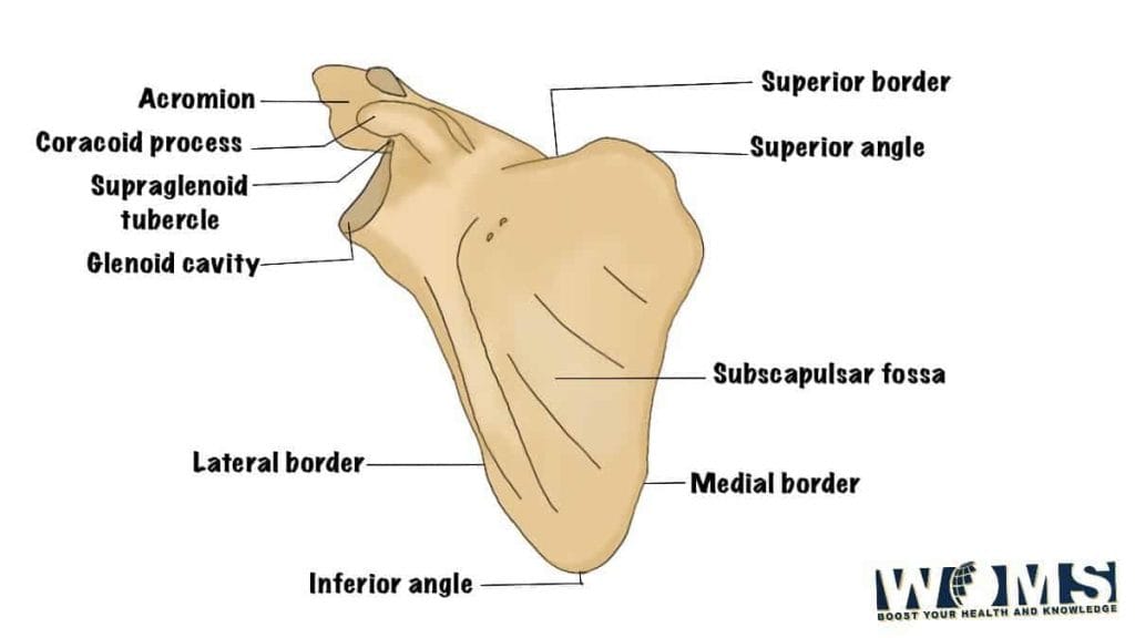
Medial Border
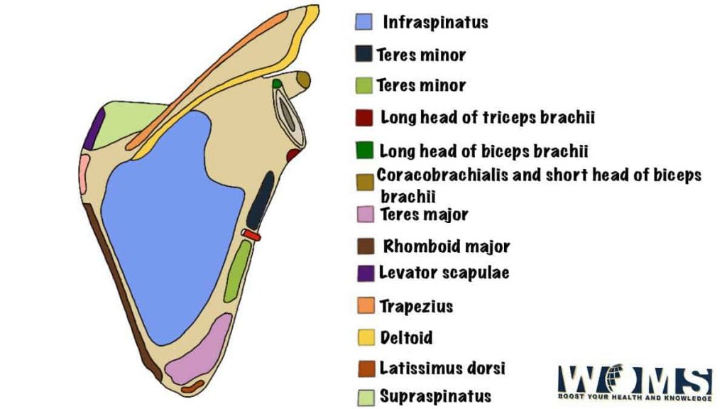
The medial border is also called the vertebral border. It is a thin border and passes closest to the spine. The medial border serves as the point of attachment of the serratus anterior on its anterior aspect and this scapular muscle performs the action of protracting and rotating the scapula.
Lateral Border
The lateral border is also called the axillary border and terminates at the lateral end of the scapula bone. It is the thickest of all the borders of the scapula. The teres minor muscle originates from the medial part of this border to cause lateral rotation of the arm.
- Glenoid cavity: It is a shallow, oval fossa that articulates 10the humerus at the glenohumeral joint.
- Supraglenoid tubercle: It is a small raised area present close to the glenoid cavity and the coracoid process.
- Infraglenoid tubercle: It is a rough elevation located inferior to the glenoid cavity.
- Supraglenoid/ greater scapular notch: The lateral border of the spine of the scapula merges with the neck of the scapula and the supraglenoid notch which bridges the supraspinous with the infraspinous fossa.
Superior Angle
The superior angle has its shape by the union of the superior and medial borders.
Inferior Angle
The inferior angle forms at the union of the medial and lateral borders and can be easily palpated. The teres major gets its origin from the posterior surface of this angle of the scapula and causes medial rotation of the arm.
Lateral Angle
At the point of fusion of the superior and lateral borders, the lateral angle comes into existence and is the broadest of all the angles of the scapula. It bears the head of the scapula.
Dorsal Surface
The dorsal or posterior surface of the scapula bone is divided unevenly into the small supraspinous and the larger infraspinous fossa by a ridge called the spine of the scapula. The supraspinatus and infraspinatus muscles occupy these fossae respectively.
The spine of the scapula
It is a triangular ridge that crosses the scapula from its medial border and merges with the acromion process laterally. The deltoid tubercle is located somewhat in the medial part of the spine and serves as an attachment for the deltoid muscle. It also provides insertion to the rhomboid major and minor muscles.
Coastal Surface
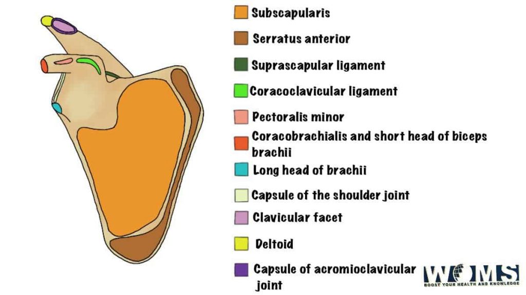
The coastal or ventral surface of the scapula rests on the posterior surface of the chest wall. It forms a large subscapular fossa that covers most of its surface. The subscapularis muscle originates from this fossa.
Acromion
The acromion (Acromian in Greek means ‘point of the shoulder’) is present as outward elongation of the spine of the scapula and is palpable is the tip of the shoulder. It forms an articular surface for the acromioclavicular joint, articulating with the lateral end of the clavicle. It provides attachment to the deltoid muscle and the trapezius muscle inserts here.
Coracoid process
The coracoid process (Greek for ‘crow’s beak’) is a broad, beak-like structure. It provides origin to the coracobrachialis and biceps brachii and insertion to the pectoralis minor and subclavius muscles. We can palpate it by applying profound pressure over the anterior surface of the deltoid muscle.
Scapular Humeral Mechanism
The scapula and the entire upper limb are suspended from the clavicle by the tense, fibrous coracoclavicular ligament strengthened by the tone of the muscles attached. The scapula bone rotates on the thoracic wall and as a result, the position of the glenoid fossa is changed, the axis of rotation now passes along the coracoclavicular ligament.
Normally, abduction of the arm involves rotation of the scapula along with the movement of the shoulder joint. At about 120 degrees of abduction, the greater tuberosity of the humeruscommunicates with the lateral end of the acromion. The further elevation is accomplished by rotating the scapula.
Clinical Relevance of Scapula
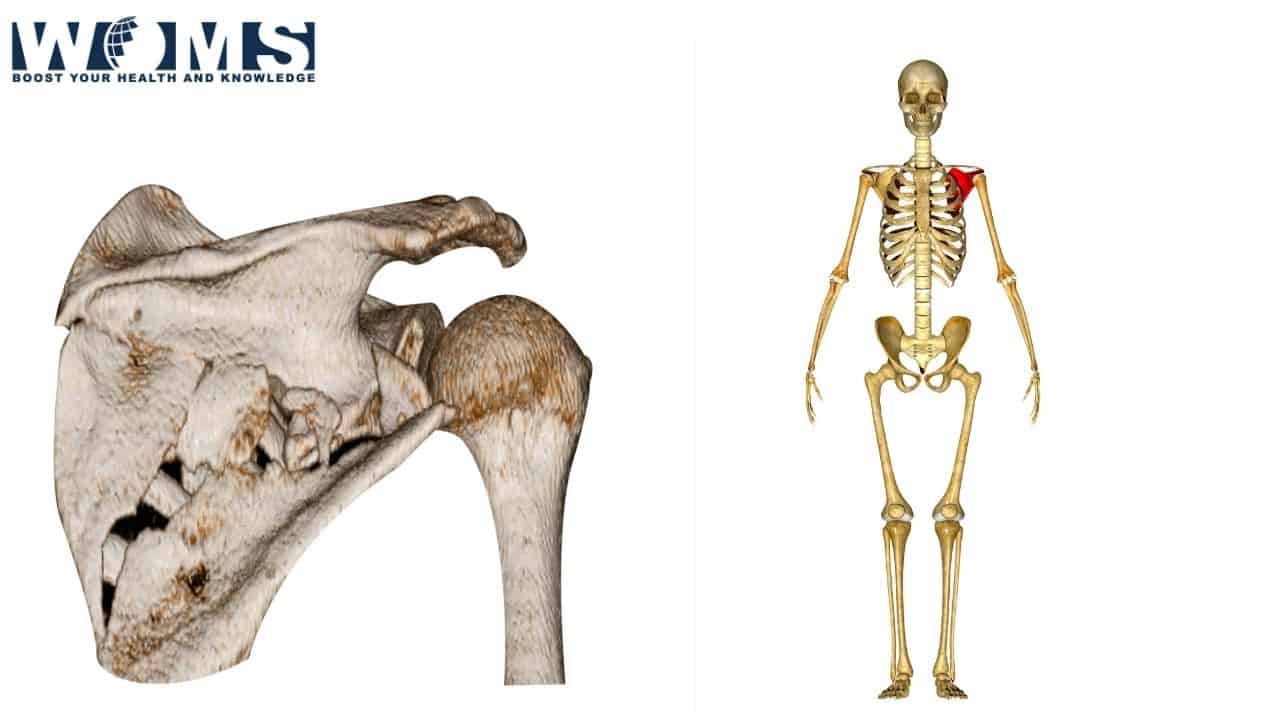
1. Scapular fractures
Scapular fractures result from severe trauma to the scapula such as in run-over accidents or pedestrian-vehicle accidents. They are usually related to fractures of the ribs.
Most scapular fractures require only a little treatment because it is supported on both sides by muscles that naturally splint the pieces together.
2. Winging of the scapula bone
The balance of the scapular muscles attached to it on both sides helps the scapula to maintain its position. If the balance is disturbed, the position becomes upset.
Injury to the long thoracic nerve paralyzes the serratus anterior muscle resulting in the scapula moving laterally and posteriorly giving the appearance of a wing. This phenomenon is a winged scapula.
The scapular winging results in failure to rotate the scapula during abduction of the arm. The long thoracic nerve may suffer injury by blows to the posterior triangle of the neck or trauma to the lateral chest wall.
How to check for winging of the scapula
A careful physical examination helps to detect it. Winging of the scapula becomes more prominent when a person leans on the wall with both hands touching the wall by the palmar aspect.
3. Triangle of auscultation
The scapula forms the lateral border of the triangle of auscultation. It is a triangular gap in the back musculature to examine the posterior aspect of the lungs with the help of a stethoscope.