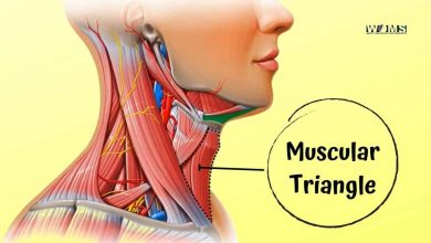
The Anterior Triangle of the Neck
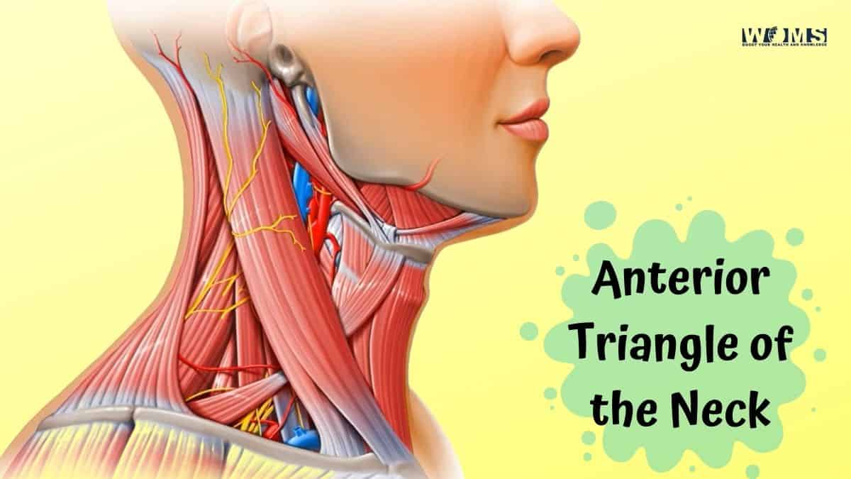
The anterior triangle of the neck is a region on the front side of your neck. It’s one of three cervical triangles, and it’s bordered by the sternocleidomastoid muscle at its base, and by the stylohyoid muscle at its apex. The anterior triangle contains numerous important structures: skin, fat, muscles (including all three scalene muscles), nerves like the cervical plexus which controls movement in your arms and hands; lymph nodes; blood vessels like the carotid artery that supplies oxygenated blood to your brain; and many others.
The anterior triangle of the neck is also a very common site for tumors and other growths. This makes it an important region to be aware of when you’re performing a physical exam on someone. It’s also important to know the anatomy of this area, in order to understand what’s going on if something seems amiss.
The anterior triangle of the neck is a space that contains important structures. It is bounded by the sternocleidomastoid muscle, hyoid bone, and cricothyroid muscle. The contents of this region are significant to head and neck surgeons as they can be injured during surgery or even by relatively minor trauma.
Structures found in the anterior triangle of the neck
Here are the structures found in the anterior triangle of the neck:
Sternocleidomastoid muscle: This large, two-headed muscle is responsible for flexion and lateral rotation of the head. It also assists in elevating the sternum (breastbone). The sternocleidomastoid muscle originates on the median plane of the manubrium (the uppermost part of the sternum) and inserts into the mastoid process of the skull.
Hyoid bone: This small, horseshoe-shaped bone is located at the base of the tongue. It plays an important role in swallowing and speech. The hyoid bone does not articulate with any other bones, but it is held in place by muscles and ligaments.
Cricothyroid muscle: This small, thin muscle is responsible for elevating the thyroid cartilage (voice box). It originates on the cricoid cartilage (a ring of cartilage at the base of the larynx) and inserts on the thyroid cartilage.
If you work in an occupation where you might experience head or neck trauma, it’s important to know how to protect your anterior triangle. The best way to do this is by wearing a cervical collar whenever possible. A cervical collar will help keep the structures in the anterior triangle stabilized and protected from injury. If a cervical collar isn’t available, be sure to protect your head and neck with adequate padding and armor if possible. By taking these precautions, you can help reduce the risk of injuries to the vital structures in the anterior triangle of the neck!
Where is the anterior neck located?
The anterior neck is located in the front of the neck. It is made up of three muscles: the sternocleidomastoid, the anterior scalene muscle, and the pectoralis minor muscle.
- The sternocleidomastoid muscle is located in the middle of the front of your neck. It runs from your breast between the mandible and sternococlemomastastoid muscle. The muscle helps to turn your head and flex the neck.
- The anterior scalene muscle is located on the side of the front of your neck. It runs from the first rib to the upper border of the scapula. The muscle helps lift your shoulder and bends your neck forward.
- The pectoralis minor muscle is located in front of your chest. It attaches to the coracoid process of the scapula and ribs three through six. The muscle pulls down on the shoulder and assists with arm movement.
Together, these muscles make up what is called the anterior triangle of the neck. This triangle extends from below your chin to just above your collarbone. It’s an important area for many reasons.
The anterior triangle contains
Superficial structures like:
cutaneous nerve, a sensory branch of cervical plexus (comes up on the outer surface) —suprascapular artery which is also a terminal branch of a subclavian artery! A really important structure coming off the axillary arch!! You don’t want it to get blocked or damaged. Can cause problems with upper limb function as well as shoulder pain etc.
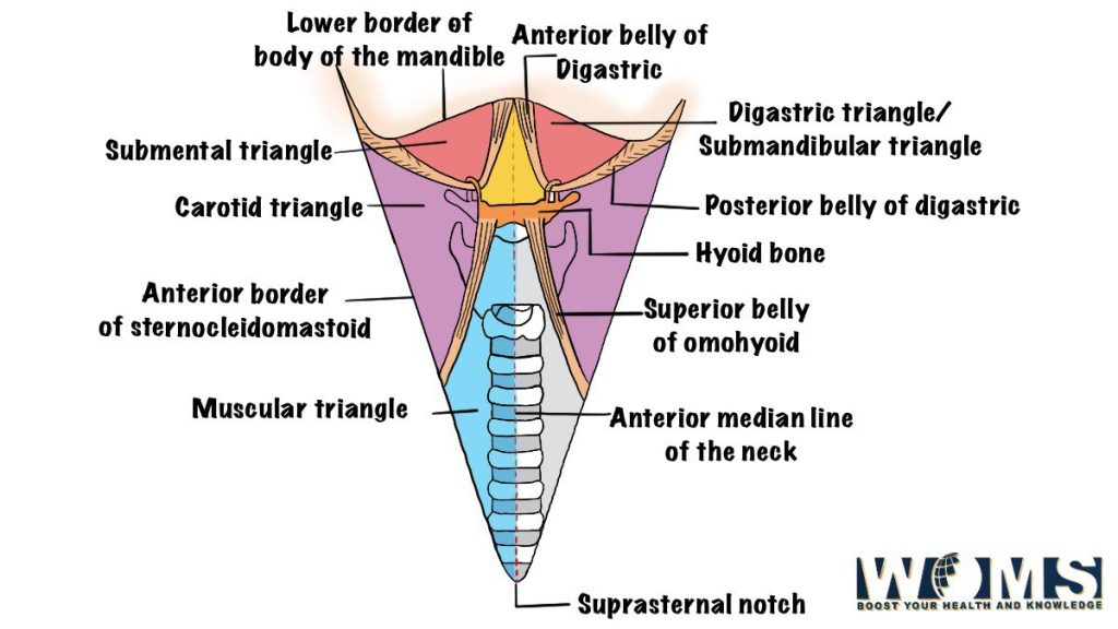
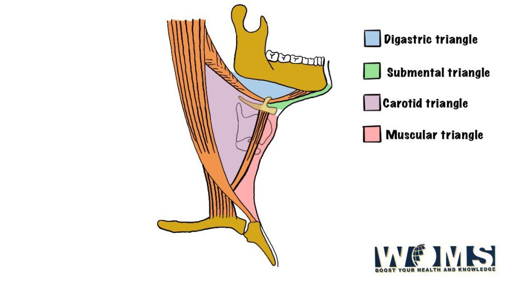
Subdivisions of anterior triangle
What Are the Anterior and Posterior Triangles of Neck?
The anterior triangle of the neck is the region bounded by the sternocleidomastoid muscle, the clavicle, and the mandible. The posterior triangle of the neck is located behind this area.
The anterior triangle includes several important structures:
- Carotid sheath, which contains the common carotid artery and internal jugular vein
- Larynx
- Thyroid gland
These structures are at risk for injury during a motor vehicle accident or other traumatic events. Knowledge of these structures is important for healthcare professionals who may be called to provide emergency care.
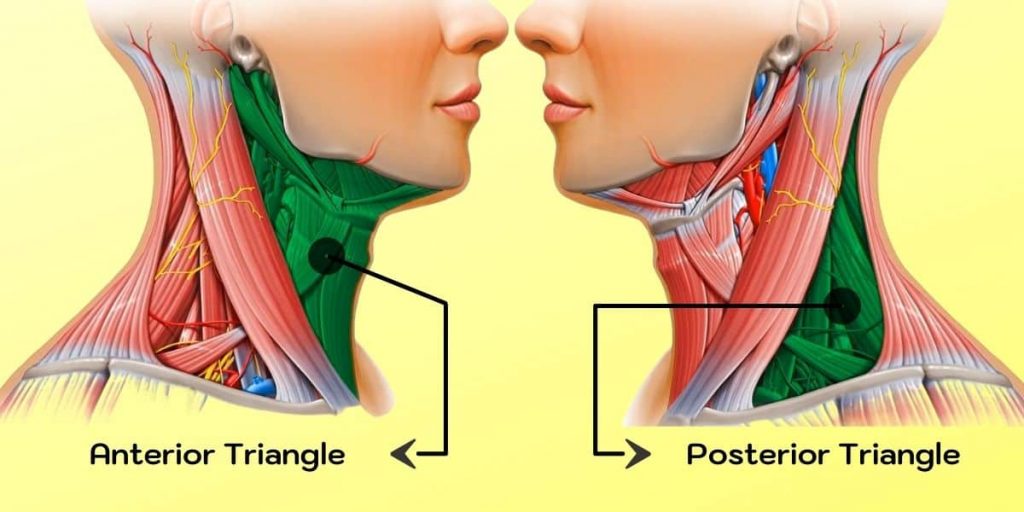
What Separates Anterior and Posterior Triangles of Neck?
The anterior and posterior triangles of the neck are separated by a muscular plane known as the “platysma” muscles that extend from the sternum to the clavicle. Another landmark includes an imaginary vertical line that runs between two points found on both sides at the top part of the trapezius visible when looking directly forward; this line represents the midpoint where lines are drawn each side from.
What Is the Importance of Anterior Triangle of the Neck?
The importance of the anterior triangle of the neck is that it contains many important structures that can be injured. These include the carotid artery, the jugular vein, and the larynx.
The carotid artery is a large blood vessel in the neck that supplies blood to the head and brain. The jugular vein carries blood from the head back to the heart. The larynx is a structure in the throat that contains the voice box.
Injury to any of these structures can cause serious problems, such as bleeding or airway obstruction. It is therefore important for everyone to know about the anterior triangle of the neck and what its boundaries are.
Boundaries of the anterior triangle of the neck
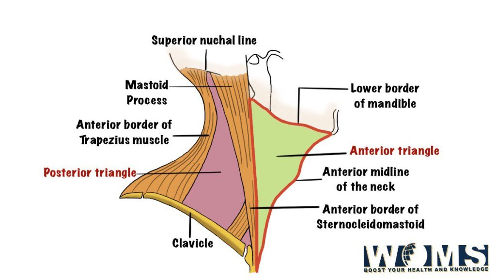
- Mandible (lower jaw) and the mastoid process (a bump on the skull)
- Middle third of the clavicle (collarbone), and a line connecting this point to both corners of the jaw.
This is where you would put your index fingers when palpating (feeling for lumps or swellings).
The base of the triangle extends from its lowest point between two lower ribs, down over nipple level, and then out toward each armpit until it reaches another boundary. This last one is:
The midline side edge of the latissimus dorsi muscle in a person’s back. This area also contains many important structures such as blood vessels and lymph nodes. It is very common for people to have pain in this region without realizing that the cause is actually something else.
Knowing about the anterior triangle of the neck and its boundaries can help you to identify any potential problems and seek medical attention if necessary. For example, if you have a lump or swelling in this area, it is important to get it checked out by a doctor. The sooner any problems are identified and treated, the better.
So now that you know all about the anterior triangle of the neck, be sure to keep it in mind next time you’re feeling lumps or swellings! And remember, if anything seems amiss, don’t hesitate to see your doctor. They will be able to tell you what’s going on and how best to treat it.
Situation of Anterior Triangle of the Neck
The anterior triangle of the neck is a region on the anterior side of the human body, where some muscles and other structures come together.
The following list gives an idea about what can be found in this area:
- Muscles- sternocleidomastoid muscle, omohyoid muscle, trapezius muscle
- Bones- clavicle bone (collarbone), scapula bones (shoulder blades)
- Veins & arteries- subclavian artery – brachiocephalic trunk
So while it might seem like a small and relatively insignificant part of your body, the anterior triangle of the neck is actually home to many important structures and should be taken into account when looking at any issues or injuries in this region.
Contents of the Anterior Triangle
The contents include several arteries and nerves along with their accompanying fascia. These structures have been divided into three groups based on embryologic development. As seen from a superior view, they have mainly related anatomically to each other rather than being involved in any functional unit.
Of course, it must be kept in mind that there may well exist considerable individual variation among the population examined during dissections at autopsy because these variations affect significant clinical implications. For example, the carotid sheath may be quite wide and deep or relatively narrow and shallow.
The Main Structures in AnteriorTriangle of the neck
The main structures are:
- Anterior jugular vein (sides)
- Anterior carotid artery (side, at the level of hyoid bone)
- Thyroid gland: The thyroid gland also lies on either side of the larynx.
- Facial nerve close to its bifurcation point.
The facial nerve gives off branches to supply muscles in parts of your face before it splits into two halves. These are known as “temporal” and “mandibular” divisions which gradually merge further down into numerous smaller groups for tiny movements like blinking or smiling that all add up together to produce recognizable expressions on our faces.
Larynx (voice box)
The larynx is a cartilaginous structure that contains the vocal cords. It sits in the midline of the anterior neck and just below it is the hyoid bone which attaches to muscles of the tongue and floor of the mouth.
The structures within the anterior triangle of the neck can be further divided into three groups:
Group I includes the muscles that form the wall of the triangle:
- Sternocleidomastoid muscle
- Omohyoid muscle
- Thyrohyoid muscle
Group II consists of blood vessels and nerves:
- Common carotid artery
- Internal jugular vein
- Vagus nerve (X)
- Accessory nerve
Why is it Important to Divide the Neck into Segments or Triangles?
It is important to divide the neck into segments or triangles because it allows us to accurately identify the structures located in each area. by defining the boundaries of these regions, we can understand how they function and interact with one another.
Applied Anatomy of the Anterior Triangle of the neck
- A goiter is an enlargement of the thyroid gland, usually caused by an iodine deficiency in the diet. It can cause a number of problems such as difficulty swallowing or breathing, hoarseness, and even changes in heart rate and blood pressure.
- The anterior triangle of the neck is an important area for many reasons and should be taken into consideration when looking at injuries or problems in this region. For example, a fracture to the clavicle bone can cause immense pain and disability as it can restrict movement of the arm. Additionally, trauma to one of the muscles in this area such as the sternocleidomastoid muscle can lead to swelling and bruising. Finally, an infection in one of the lymph nodes present here can cause a condition known as Ludwig’s Angina which results in severe difficulty swallowing and potentially even death if not treated promptly.
Also read: Submental Triangle
Conclusion
The anterior triangle of the neck is a region in the upper part of the human body, situated roughly at its center. It includes several structures including muscles and bones. Anatomists typically use landmarks on three sides of this area to describe it: beginning with one corner formed by points between chin and sternum (the jugular notch), passing through two more corners made up from where the jawbone meets earlobe, then down alongside the neck’s edge to meet clavicle bone just past the point where collarbones diverge slightly. The line that runs across these points can be considered an imaginary boundary for this anatomical structure.
FAQs
What is the anterior triangle of the neck?
The anterior triangle of the neck is a region on the front of the neck. It is bounded by three lines:
1. The midline of the neck, which runs from the chin to the top of the chest
2. The lateral line, which runs along each side of the neck from just in front of the ear to just behind the shoulder
3. The inferior border, which curves around from just below the ear to just above the clavicle (collarbone)
What muscles are contained within it?
The muscles contained within this area include:
1. Sternocleidomastoid muscle
2. Scalene muscles
3. Thyroid gland
What is its function?
The main functions of this area are:
1. To allow movement of the head – The muscles within the triangle provide a lot of flexibility for movements such as nodding or tilting your head back
2. To protect underlying structures – The triangle contains many important blood vessels and nerves which run up into the head.