Anatomy of Abdomen
The abdomen is also known as the belly. It is a body space situated between the thorax and the pelvis. Knowing about the anatomy of abdomen will give us an in-depth understanding of what our abdomen is like. Let’s learn more about the anatomy of the abdomen.
The abdomen is the part of the trunk which lies just below the diaphragm. It is a cylindrical chamber extending from the inferior margin of the thorax to the superior margin of the pelvis and lower limb.
The abdomen is margined superiorly by the inferior thoracic aperture and inferiorly by a pelvic inlet which forms two-cavity where the upper part is known as the abdomen proper and the Lower part is known as the pelvic cavity.
Viscera of abdomen
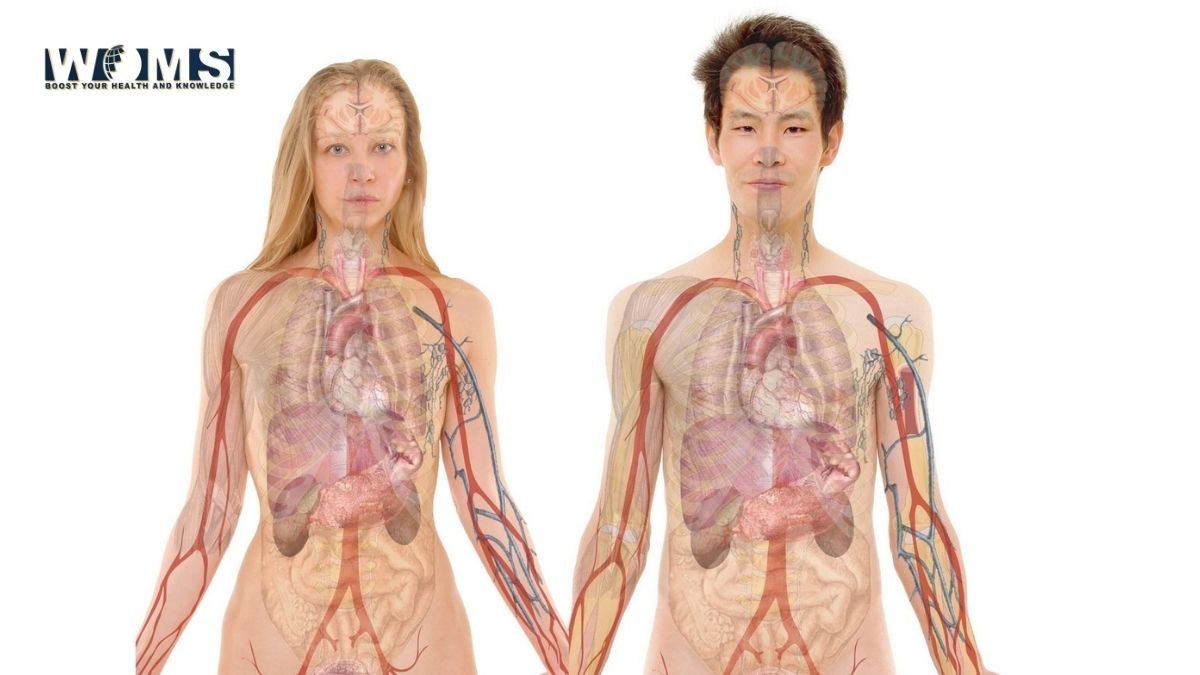
Abdominal viscera are suspended in the peritoneal cavity by the mesenteries. It is positioned between the cavity and the musculoskeletal wall. Abdominal anatomy includes a major element of the gastrointestinal, system, the caudal end of the esophagus, stomach, large and small intestine, liver, pancreas, and gallbladder. It also contains the spleen, the component of the urinary system, kidney, and ureter.
Furthermore, it contains the suprarenal glands, and the major neurovascular structure is present in the abdominal viscera. The anatomy of abdomen also tells about the viscera of the abdomen.
Functions of the abdomen
- It acts as a house protecting the viscera like the gastrointestinal tract, esophagus, liver, intestine (large and small), gall bladder, and stomach.
- It also helps in breathing and changing the intraabdominal pressure by contraction of the abdominal wall.
Boundaries of abdomen
The anatomy of abdomen tells us about its boundaries as well; Roof, floor, anterior wall, and posterior wall from the boundary of the abdomen.
Roof: – undersurface of the diaphragm from the roof of the abdomen which moves up and down during breathing and changes the abdominal pressure.
Floor: – pelvic diaphragm from the floor of the pelvic cavity in the posterior part and the urogenital diaphragm forms the floor of the pelvic cavity in the anterior part.
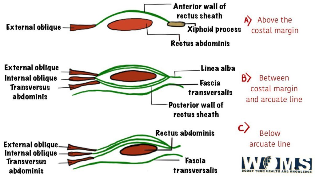
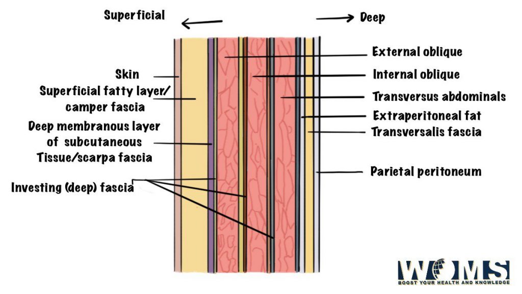
Anterior wall: – It is formed by the three muscles which are flat and by their aponeuroses. They are rectus abdomens and pyramidal muscles within the rectus sheath. The anterior wall is elastic & firm which protects the abdominal viscera
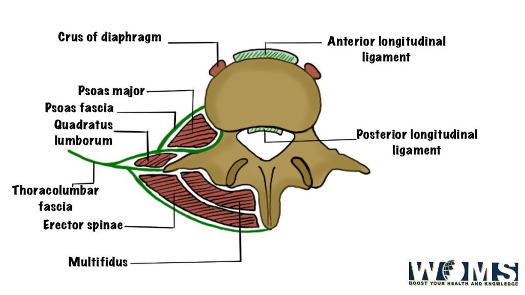
Posterior wall: – various retroperitoneal organs, main vessels and nerves are present in the posterior wall. It provides support & nutrition to the various organs by the attachment of various peritoneal folds.
Regions of abdomen
What do we mean by abdominal cavity?
Let’s see the anatomy of the abdomen to know about the abdominal cavity. The larger upper part is known as the abdominal cavity which is subdivided by the plane of the Pelvic inlet.
It can also be called the abdominal cavity proper. The abdominal cavity proper is mainly used as a clinical term.
It is the largest cavity of the body. The boundaries of the abdominal cavity are defined below:
- Superiorly: Diaphragm, which separates it from the thoracic cavity
- Anteriorly: Anterior abdominal wall, abdominal muscles
- Posteriorly: Posterior abdominal wall, formed by lumbar.
- Inferiorly: Continues with the pelvic cavity at the pelvic inlet.
- Laterally: muscles and Lower rib of the Anterior Abdominal wall
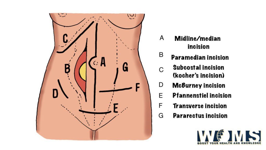
Abdominal incisions
What is the content of the abdominal cavity?
The anatomy of the abdomen also deals with the contents present in the abdominal cavity. Moreover, the content of the abdominal cavity is the organs and glands of the digestive and urinary systems which occupy most of the part of the abdominal cavity.
The anatomy of abdomen includes; the anatomy of the liver, anatomy of the small intestine, anatomy of the large intestine, anatomy of the prostate, anatomy of the vagina, anatomy of the seminal vesicles, and the anatomy of the uterus, the anatomy of ureter and more. These organs and glands of abdominal anatomy are present below:
- Stomach, small intestine, and most of the large intestine.
- Liver, gallbladder, and pancreas.
- Two kidneys and the upper part of the ureters.
- Adrenal glands which are also known as suprarenal glands
- 50ther structures include blood vessels, lymph vessels, nerves, spleen, and lymph nodes.
Stomach
One of the major viscera of the abdominal cavity is the stomach. Here you can learn the anatomy of the stomach with its function
The stomach is one of the largest parts of the digestive tube. Its shape is just like an alphabet “J” and is located in the abdominal cavity just below the Diaphragm, a little bit left of the midline. An adult has an average volume capacity of about 1500ml.
Functions of stomach
The major functions and activities carried out by the stomach are as follows:
- Churning and breaking of food and mixing it with the Gastric juice secreted by specialized glands in its mucosa.
- Storing the food temporarily.
- Secreting intrinsic factor needed for absorption of Vitamin B12.
Small intestine
The small intestine is a convoluted tube connecting the stomach with the large intestine. It is about 6 m in length and extends from the pyloric sphincter to the ileocecal junction. However, the small intestine is present in the central part and lower parts of the abdominal proper cavity where the large intestine surrounds it.
The three parts of the small intestine are:
- Duodenum
- Jejunum, and
- Ileum.
The duodenum is present side by side and is a short curve (c-shape) portion and is about 25 cm long. It is the wider part of the small intestine. The ducts from the gallbladder and liver and pancreas enter the duodenum of the small intestine.
Furthermore, the jejunum is the remaining upper two fifth parts of the small intestine and the lower three-fifth is termed the ileum.
The main functions of the small intestine are digestion of food and absorption of nutrients.
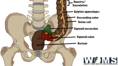
Large intestine
The large intestine is one of the most significant parts of the abdominal cavity. Many organs are present in the anatomy of the large intestine, which are present below with their descriptions.
The large intestine starts at the end of the ileum as the caecum and ends at the anus. Its length is about 1.5 m long and forms an arch coiled around the small intestine. The large intestine can be described as it is divided into the following seven parts:
- Caecum and appendix.
- Ascending colon.
- Transverse colon.
- Descending colon.
- Sigmoid colon.
- Rectum.
- Anal canal
Functions of the large intestine
The main functions of the large intestine are as follows:
- It helps in the Absorption of water and salts.
- It helps in the Formation and excretion of feces.
Liver
The liver is one of the largest glands present in the abdominal cavity of the body.
Where is the liver located?
It is present in the upper right part of the abdominal cavity with two major lobes right and left. The right lobe is much larger than the left lobe. Dealing with its location, and function includes all detail about the anatomy of the liver.
Functions of the liver
Some important functions of the liver are as follows:
- Helps in the Metabolism of carbohydrates, fat, and protein.
- Helps in the detoxification of drugs and poisons.
- It also helps in the Storage of glycogen and fat-soluble vitamins (ADEK).
- Secretion of bile.
Gallbladder
The anatomy of the abdomen includes the anatomy of the gallbladder too. It has a shape like a pear.
In addition, it is present on the inferior surface to the right lobe of the liver. It receives bile secreted by the liver, which it stores and concentrates. When fatty food enters the duodenum, the bile pours into the intestine through the bile duct by the contraction of the walls of the bladder.
- Jaundice is a clinical condition that has characteristics like yellowing of the skin and sclera of the eyes. This condition occurs when bile enters the blood in liver diseases such as hepatitis or cirrhosis of the liver where the liver cells break down and release bile into the blood
Pancreas
The pancreas is an elongated lobulated and soft pale gland; it is a finely lobulated gland with a grey color. However, it is about 12–15 cm long and situated transversely across the posterior abdominal wall. It has got a broad head, neck, body, and a narrow tail.
Let’s start anatomy of the pancreas with its head. The Head of the gland is present within the duodenum curve, the body is present behind the stomach and the tail is present in front of the left kidney. The tail goes as far as to the spleen.
Functions of pancreas
- It is an exo-endocrine gland. The function of the exocrine part of the pancreas is to produce pancreatic juice containing enzymes which help in digestion of carbohydrates, proteins, and fats.
- The function of the endocrine pancreas is to secrete hormones, insulin and glucagon, which control the blood glucose level.
Clinical note for the pancreas
- The Deficiency of insulin released by pancreas results in diabetes mellitus.
Spleen
The spleen is a large wedge-shaped mass of vascular and lymphoid tissue. It is purplish-red and is present high up at the back of the abdominal cavity on the left side behind the stomach.
Functions of spleen
The main functions of the spleen are as follows:
- Destruction of red blood cells.
- Production of fresh lymphocytes for the bloodstream.
This is all about the anatomy of the spleen with its functions.
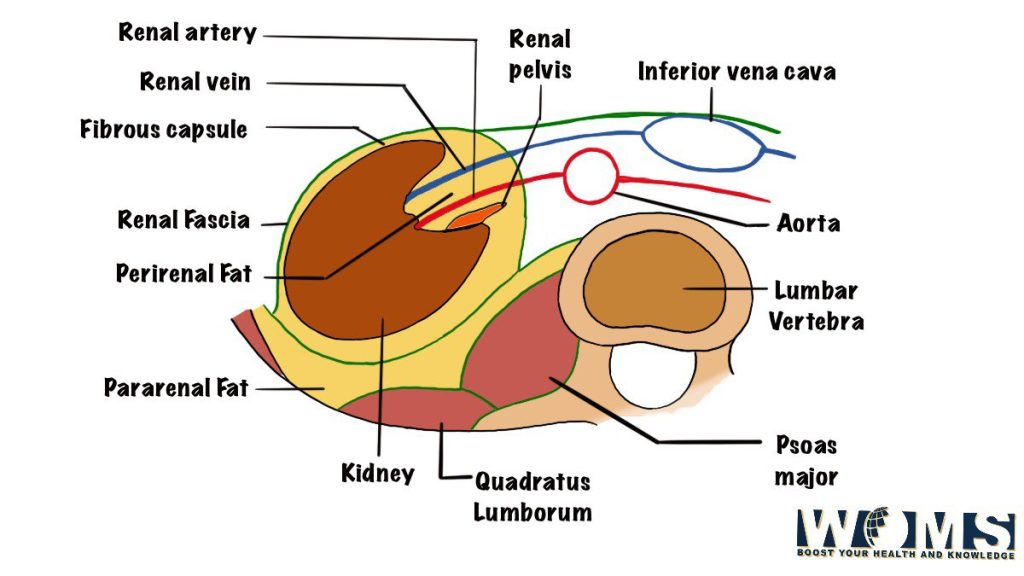
Kidneys
The anatomy of the kidney begins with its shape and location. It usually has the shape of a bean.
where are the kidneys located? Also do read the detail about the anatomy of kidney, the anatomy of the ureter, and its location.
It is present in pairs and it is an organ present on the posterior abdominal wall, one on each side of the vertebral column, behind the peritoneum. The right kidney is present usually at a slightly lower level than the left kidney.
Functions of kidneys
- Helps to secrete and excrete urine.
Ureters
The ureter is part of the pelvic cavity there are two tubes through which connect the kidneys to the urinary bladder. Both ureters are about 25 cm in length and having a diameter of about 3 mm.
Functions of ureters
- Ureters help in the transportation of urine from the kidneys to the urinary bladder.
Adrenal glands or Suprarenal glands
It is also one of the important glands of the abdominal cavity. Two suprarenal glands are the right and left adrenal glands. Both glands cover the upper pole of the corresponding kidney.
The right gland is triangular whereas the left gland is semilunar (half moon-like) in shape. Each gland consists of two parts: They are cortex and medulla.
The function of the cortex is to secrete several steroid hormones, which are responsible for:
- Maintenance of electrolyte and water balance,
- Maintenance of blood sugar concentration and of the liver and muscle glycogen,
- Control of inflammatory reactions.
The function of the medulla is to secrete adrenaline and non-adrenaline in the blood, which serve as neurotransmitters. Check the next post for details on the anatomy of the adrenal gland. You can read it in the post on the anatomy of the kidney.
Urinary Bladder
The urinary bladder is part of the pelvic cavity. It acts as a reservoir of urine. Its size, shape, and position vary with the amount of urine present there.
When it is empty it lies in the pelvic cavity, but when it distends with the urine it expands upward and forward into the abdominal cavity. The bladder can hold normally up to 500 ml to 600 ml of urine, though this causes pain and also may lead to severe anomalies in the future.
However, the urge to urinate is usually felt when it contains around 250–300 ml of urine. The two ureters enter into it through its posterior wall.
Furthermore, the urethra leaves the bladder at its neck. The bladder has got neck which is the lowest and most fixed part of the urinary bladder. A thickening of smooth muscle called the internal urethral sphincter surrounds the opening of the urethra. It’s all about the anatomy of the urinary bladder and the functions of the urinary bladder are present below.
The function of urinary bladder
- The urinary bladder stores urine and passes it into the urethra.
Urethra
The urethra is a narrow canal. It extends from the inner urethral orifice in the urinary bladder to the outer urethral orifice. In a male, the urethra length is about 18–20 cm long and in a female, the length of the urethra is about 4 cm long.
Function of Urethra
In a male, the urethra serves as a common passage for both semen and urine whereas, in a female, the urethra discharges urine from the body.
Prostate Gland
The prostate is fibrous and the Muscular glands commonly known as fibro-musculoglandular organ, helps to surround the commencement of the urethra in the male.
It is present in the pelvic cavity behind the pubic symphysis and in front of the rectum. The size of the prostate gland is about chestnut and the urethra and ejaculatory ducts transverse it.
The function of the prostate gland
The function of the prostate is to secrete a thin, milky fluid, which forms about 30% of the semen and gives it a milky appearance. The prostatic secretion is alkali in nature and provides nourishment to the sperms.
Seminal vesicles
The seminal vesicles are two small fibromuscular pouches lying between the base of the bladder and the rectum
The function of seminal vesicles
The seminal vesicles secrete an alkali-containing fluid which also contains nutrients for the sperms and forms about 60% of the seminal fluid.
Ejaculatory Ducts
In the title anatomy of the abdomen, you must also read the detail about the anatomy of ejaculatory ducts. The ejaculatory ducts are two narrow tubes about 1 cm in length.
Both the duct of the seminal vesicle and ductus deferens unite to form the ejaculatory duct. They traverse the prostate gland to open into the urethra within the gland (prostatic part of the urethra).
Functions of Ejaculatory ducts
The ejaculatory ducts pass the secretions of testes and seminal vesicles into the prostatic urethra.
Uterus
The uterus is hollow; has a thick wall present and the muscular organ is present in the pelvic cavity in between the urinary bladder and the rectum.
Moreover, the anatomy of the uterus mainly deals with its shape because shapes really matter for any disease conditions and other issues. Its shape looks like a pear and it’s anteroposteriorly is flat.
In the majority of women, the uterus leans forward (anteflexion) and bends forward (anteversion) almost at a right angle to the vagina. In the erect posture, the uterus lies in an almost horizontal position.
The uterus gets divided from above down into the following three parts:
- fundus,
- body, and
- cervix.
The cervix moves into the vagina through its anterior wall and reaches into it at the external OS. On either side, it communicates with the uterine tubes, which open into it. Below it communicates with the vagina
Functions of uterus
The functions of the uterus after puberty are:
- It is Responsible for the menstrual cycle (MC).
- Provides implantation to zygote in its wall.
- It allows the growth and development of the embryo and fetus.
Vagina
The vagina is a fibrous and muscular tube known as fibromuscular which extends obliquely downward and forward from the uterus to the vestibule of the vagina the elliptical space between the labia minor and labia major.
It is located in between the urinary bladder from the front side, and the rectum and anal canal behind. The vagina has no secretory glands though it is moist by the various cervical secretions.
Functions of vagina
The functions of the vagina are as follows:
- It acts as a receptacle for the penis during copulation.
- It gives an elastic passage to during delivery for the birth of the child.
Uterine tubes
There are two uterine tubes. Both tubes are about 10 cm in length and extend from the side of the uterus which means between the fundus and body to the ovary where it opens into the peritoneal cavity.
It gets divided into the following parts from the distal to the proximal end:
- Infundibulum,
- Ampulla,
- Isthmus, and
- Intramural.
Functions of uterine tubes
The functions of uterine tubes are as follows:
- It helps to Transport the ovum from the ovary to its lumen and provides a proper suitable site for fertilization.
- It helps in the transportation of the fertilized ovum to the uterine cavity for implantation.
Ovaries
The ovaries are female gonads. Each ovary is about the size and shape of an almond and is situated in a shallow fossa on the lateral wall of the pelvic cavity behind and below the distal end of the uterine tube.
Functions of ovaries
The ovaries start their function at puberty and continue to discharge the ova every month from about the age of 13 years old to the age of 45 years. For now, we are done discussing all the detail of the anatomy of the abdomen. Now you need to know about the regions of the abdomen.
Regions of the abdomen
The anatomy of abdomen also tells about the regions of the abdomen. Moreover, the region of the abdomen is also known as the quadrant of the abdomen.
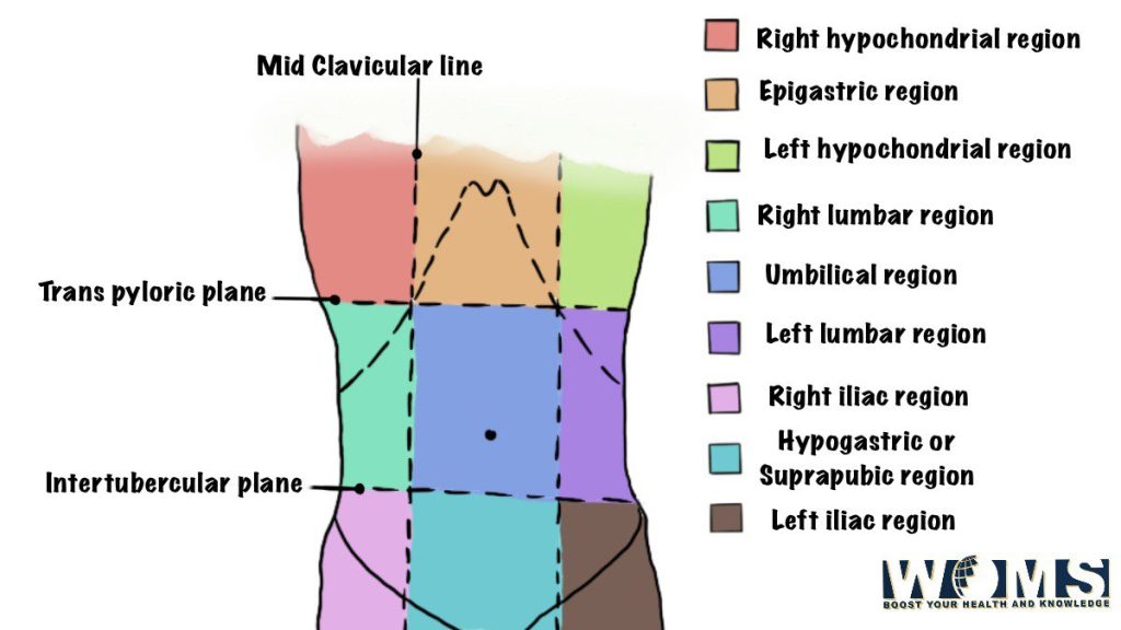
The abdominal cavity is conventionally divided by the clinicians into nine regions by two horizontal and two vertical lines. A Vertical line pass through the mid-inguinal point and the horizontal passes by the Upper horizontal line corresponds to the transpyloric plane. The lower horizontal line between the tubercle of the Iliac crest.
The nine regions of the abdomen are:
- Epigastric: Liver, stomach, pancreas, duodenum lies in this region
- Left Hypochondriac Region: Spleen, colon, liver, left kidney, small intestine lies in this region
- Right Hypochondriac Region: Gallbladder, liver, right kidney lies in this region.
- Umbilical Region: Naval, duodenum, colon, kidneys, major abdominal vessels lies in this region
- Left Lumbar Region: Descending colon, left kidney, small intestine lies in this region.
- Right Lumbar Region: Cecum, ascending colon, liver, right kidney, small intestine lies in this region.
- Hypogastric Region: Urinary bladder, small intestine, sigmoid colon, female reproductive organs lies in this region.
- Left Iliac/Inguinal Region: Small intestine, descending colon, sigmoid colonies in this region.
- Right Iliac/Inguinal Region: Appendix, cecum, small intestine lies in this region.
- Hernia: protrusion of an organ through the wall, which retains it. The common abdominal hernias are inguinal, umbilical, femoral, and incisional.
- Abdominal pain: intra-abdominal diseases are Present with pain only. The two important features of abdominal pain are its site and its nature. Clinician mainly diagnoses by having a clear idea of the region where there is pain.
- Abdominal mass: The common cause of abdominal Masses are: hepatomegaly where the liver enlarges. Splenomegaly where the spleen enlarges, hydronephrosis where the kidney enlarges, and pseudocyst of the pancreas.
If you are aware of the anatomy of the abdomen then you can distinguish the disorders present in the individual.
Abdomen Conditions
1. Constipation
Having fewer than three bowel motions each week is constipation. Diet and exercise may be beneficial, but many people will need to consult their doctors.
2. Gastritis
It is a stomach inflammation that can cause nausea and/or pain. Gastritis can be caused by a variety of reasons, including alcohol, NSAIDs, H. pylori infection, and others.
3. Peptic ulcer disease (PUD)
It is a type of stomach ulcer. It refers to acid, and ulcers are erosions. Peptic ulcers are stomach and duodenal ulcers (the first part of the small intestine). An infection with H. pylori or the use of anti-inflammatory medicines such as ibuprofen is the most common cause.
4. Peritonitis
It is an inflammatory condition that affects the covering of the abdominal organs, resulting in abdominal wall stiffness and severe pain. This is usually caused by a burst or an infected abdominal organ.
5. Acute abdomen
It is a medical term that doctors use to indicate the presence of peritonitis or another emergency that necessitates surgery.
6. Appendicitis
It is a condition in which the appendix in the lower right colon becomes inflamed. An inflamed appendix is usually surgically removed (appendectomy).
6. Cholecystitis
It is a gallbladder inflammation that causes significant right-sided abdominal pain. The most common cause is a gallstone blocking the duct exiting the gallbladder.
7. Pancreatitis
It is a condition in which the pancreas is inflamed. The most prevalent causes of pancreatitis are alcohol and gallstones. Other factors include drugs and trauma; around 10% to 15% of instances are caused by unknown factors.
8. Hepatitis
It is an inflammation of the liver caused by a virus. Hepatitis can also be caused by drugs, alcohol, or immune system disorders.
9. Cirrhosis
It is a condition in which the liver scars as a result of chronic inflammation. The most common causes are excessive drinking or chronic hepatitis.
Cirrhosis is a common cause of ascites, which is a buildup of fluid in the abdomen. Ascites can make the abdomen protrude noticeably.
Takeaway:
The abdomen is the part of the trunk which lies just below the diaphragm. It is a cylindrical chamber extending from the inferior margin of the thorax to the superior margin of the pelvis and lower limb. It is margined superiorly by the inferior thoracic aperture and inferiorly by a pelvic inlet which forms two-cavity where the upper part is known as the abdomen proper and the Lower part is known as the pelvic cavity.
I hope after reading this you have an idea about the anatomy of the abdomen. I hope if this article helped you to know more about the anatomy of the abdomen do share it.
Additional Images
FAQs:
1. What are the types of abdomen pain?
There are three types of abdomen pain. They are visceral pain, referred pain, and parietal pain.
2. What is the anatomy of the abdomen?
The abdomen is also called the belly. It comprises the area that is present between the thorax and pelvis. Anatomy of abdomen comprises from the area, organs present in the abdomen to the blood vessels and nerve supply.
3. How trapped gas does feel like?
When you have trapped gas then you may feel a stabbing pain in your chest or abdomen. This stabbing pain can be so sharp that you might feel that it is a heart attack, appendicitis, or gallbladder problem.
4. How does abdominal pain last?
If it is normal pain and has no underlying problem then it might subdue after two hours.
5. What is the main difference between the cephalothorax and abdomen?
The cephalothorax is the region of the body that is formed by the fusion of the head and the thorax. It is present in the Chelicerates and crustaceans of the arthropods group.
While abdomen is the part of the body that is located posterior to the thorax. It is present in the animals who have evolved after annelids and arthropods.