Unveiling 6 Secrets of Ferret Skull Anatomy and its Importance in Veterinary Medicine
Ferret is a domesticated species belonging to the class Mammalia. It belongs to the Mustelidae family. In appearance, ferrets resemble other mustelids because of their slender and long bodies. This article provides detailed information regarding the ferret skull and associated structures. In addition, it will help the veterinary students to learn about the anatomical aspects of the ferret skull in detail.
The ferret skull is flat and elongated with a small facial region. The facial region is about one-third of the length of the cranial part. Moreover, the adult skull is longer and wider than its female counterparts. The ferret skull anatomy is divided into six standard views to illustrate all the important bony structures in detail. These six views are dorsal, ventral, lateral, caudal, cranial, and midsagittal. The mandible is a separate bone projected from the cranium. Let us have a detailed look at the morphological aspects of the ferret skull.
1. Dorsal view of the ferret skull
The dorsal view of the ferret skull exhibits different important structures in the cranial and facial portions. Let us have a look at the cranial and facial portions to illustrate the anatomical details of the dorsal view.
Cranial portion
The most important and visible structure on the dorsum of the cranium is the external sagittal crest. It is made up of different bones including frontal, parietal, and interparietal bones. This structure is more prominent in male ferrets than in females. In a rostral direction, the crest is divided into a ‘V’ form, extending towards the postorbital processes of the frontal bone on both sides of the skull. This crest starts to divide earlier into females rather than males.
The differentiating structure between the dorsal and caudal view is the transverse crust or nuchal crest. It is formed by the parietal and occipital bones. Moreover, the dorsum of the ferret skull exhibits a convex and broad surface that demarcates the temporal fossa. It is the area from where temporal muscle arises.
Facial portion
The facial part of the ferret skull is quite short in the dorsal view. It consists of the dorsal aspects of maxillary, nasal, premaxillary bones, and nasal processes of the frontal bone. The premaxillae form the most anterior margin of the upper jaw. In addition, it makes the border of the external nasal opening combine with the nasal bones.
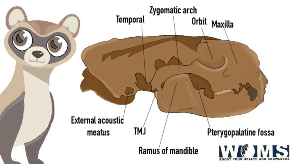
2. Lateral view of the ferret skull
Cranial portion
In the lateral view, the most prominent structures are the orbit and projecting zygomatic arch. The zygomatic arch curves in the dorsal and lateral directions. In addition, it bends from the zygomatic process of the maxilla towards the zygomatic process of the temporal bone. In this way, it forms a de3p large cavity on both sides. The zygomatic arch performs three main functions. These are as follows:
- Serving as an origin point for the masseter and a small part of the temporal muscle
- Supporting the eye
- Offers an articulation point for the mandible
The orbit is the continuation of the ventrally located pterygopalatine fossa due to the absence of a postorbital bar. In the medial aspect, the ventral orbital crest demarcates the border of the two fossae. In addition, it also marks the boundary for the origin of the medial pterygoid muscle.
Facial portion
In the lateral view, the maxilla is the most important structure dominating over any other bone. The infraorbital foramen lies ventrally to the lower orbital margin facing the rostrolateral direction. The alveolar jugum of the canine is also visible in the lateral view of the ferret skull.
3. Ventral view of the ferret skull:
Cranial portion
The ventral view of the cranium explores the tympanic bullae and occipital condyles. The tympanic bullae are two prominent protuberances on both sides of the ventrocaudal aspect of the cranium. Medially to the tympanic bullae, there are basioccipital, basisphenoid, and presphenoid bones. The basioccipital bones fuse in a lateral direction to form the tympanic and petrous parts of the temporal bone. In addition, it articulates with the body of the basisphenoid rostrally. The basisphenoid bones combine with the pterygoid and presphenoid bones rostrally. Laterally, the presphenoid bone connects with the perpendicular part of the palatine bone.
The mandibular fossa is also visible as a transverse groove in the ventral view. A small post-articular process projects from the mediocaudal basal part of the mandibular fossa. It works as a posterior bracing point for the mandibular condyles.
Facial region
The ventral view of the facial region depicts the teeth and hard palate. There are three incisors in the premaxilla region. In a posterolateral direction to the maxilla, there are one upper canine, three premolars, and one molar. The horizontal portion of the premaxilla, maxillae, and palatines form the base of the hard palate. The hard palate extends up to the posterior margins of the molars. A small ventral opening of the incisive canal is present in the pre-maxillary region. In this view, the major posterior palatine foramina are visible and present medially to the third premolars.
4. Caudal view of the ferret skull:
The occipital bone is the main finding in the caudal view of the skull. It consists of paired exoccipitals with occipital condyles, supraoccipital, and notches basioccipital portion. The nuchal crests are formed by the lateral borders to meet the squamous and parietal parts of the temporal bones. The nuchal crest intersects the external sagittal crest by concaving rostrally at the mid-dorsal point. In the caudal view, the mastoid process is clearly visible and well-developed. It combines with the ventral end of the nuchal crest. The foramen Magnum is also visible at the ventral area of the occipital bone.
5. Rostral view of the ferret skull
The rostral or frontal aspect is made up of the frontal ends of the upper and lower jaws. There are six incisors and two canines in both jaws. There is also a diastema present lateral to the upper third incisor on both sides. This diastema provides a pathway for the lower canine to come into occlusion. The external nasal openings are also visible through which ventral and dorsal nasal conchae are seen.
6. Midsagittal view of the ferret skull
The nasal cavities are prominent in the midsagittal view. It consists of two symmetrical halves separated by the nasal septum. Every half consists of ventral nasal conchae (ventro-rostrally), and delicate turbinates of the ethmoid (dorsocaudally). There is also a maxillary sinus present over the hard palate. The maxillary sinus moves laterally and caudally. A thin lateral wall separates the roots of the upper 3rd and 4th premolars from the maxillary sinus. The frontal and sphenoid sinuses are also visible in this view. At the caudal end, the cribriform plate of ethmoids separates the nasal cavity from the cranial cavity.
Cranial cavity
The cranial cavity is composed of three main sections. The olfactory fossa is present caudally to the cribriform plate and contains olfactory bulbs. In continuation to the olfactory fossa, there is the extensive cerebral fossa posteriorly to house the cerebrum, diencephalon, and mesencephalon. In the caudal end of the fossa, there is a hypophyseal fossa. The tentorium marks the termination point of the cerebral fossa. There is a cerebellar fossa posterior to the tentorium . It surrounds the pons, cerebellum, and medulla oblongata.
There is a small bird-shaped bony structure that is present on the top of the petrosum, anterior to the internal auditory meatus, characterizing the ferret skull in the sample.
Mandible
The mandible is made up of a pair of dentary bones that articulate with the mandibular symphysis anteriorly. From the symphysis point, these two dentary bones diverge from each other to form a V-shaped mandibular space. The body of the mandible contains lower teeth. There is also a diastema between the canine and 2nr premolars and between the 3rd and 4th premolars. The body of the mandible is smooth and contains multiple mental foramina. The largest mental foramen is located ventral to the septum between the 4th and 4th premolar. The ramus of the mandible looks like a triangle and consists of three noticeable processes.
The condylar process is present at the level of posterior teeth and expands transversely articulating with the apex of the cone. It forms the temporomandibular joint by communicating with the temporal bone.
What are the general differences between male and female ferret skulls?
There are some differences between male and female ferret skulls. These are as follows:
- The male ferret skull is 17%, 22%, and 12% larger than the female skull in the context of skull length, maximum skull width, and minimum frontal width respectively.
- The variations in measurements for male skulls are greater than for females.
- The proportion of the maximum skull width to skull length is greatly higher for males than females. A male ferret skull is wider than a female ferret skull.
- The minimum frontal width in females is more than the males.
Conclusion
The ferret skull is an elongated and flat but small facial region. The typical shape of the ferret skull performs the main role of housing the brain, food, and respiratory tracts. The short facial length in the ferret skull enhances the biting strength at the front region of the jaws. In addition, the ferret skull is studied from six different views to understand the basic anatomical details related to the ferret skull. Give this article a thorough read to properly understand the basic concepts related to the ferret skull.
Frequently asked questions (FAQs)
What are the proper dimensions of the ferret skull?
The average ferret skull length is 6.5cm, the average skull width is 3.7cm, and the average skull height is 3.0 cm.
How many bones are part of the ferret skeleton?
The ferret skeleton consists of almost 200 bones. These bones are fragile and brittle so they can easily fracture.