Posterior Tibial Artery
The posterior tibial artery is a vessel of the human leg and is a large and more terminal branch of the popliteal artery. It contributes towards supplying blood to the posterior compartment of the human leg along with the popliteal artery and to the foot by its terminal branches. It gives off eight branches; two of which are terminal branches called the medial and lateral plantar arteries.
The course of posterior tibial artery
It starts its course between the bones, tibia, and fibula, at the distal end of the popliteus. It arises from the popliteal artery which is the parent artery as the former passes beneath the soleus and gastrocnemius muscles. The posterior tibial artery gives off the fibular artery which is a larger branch.
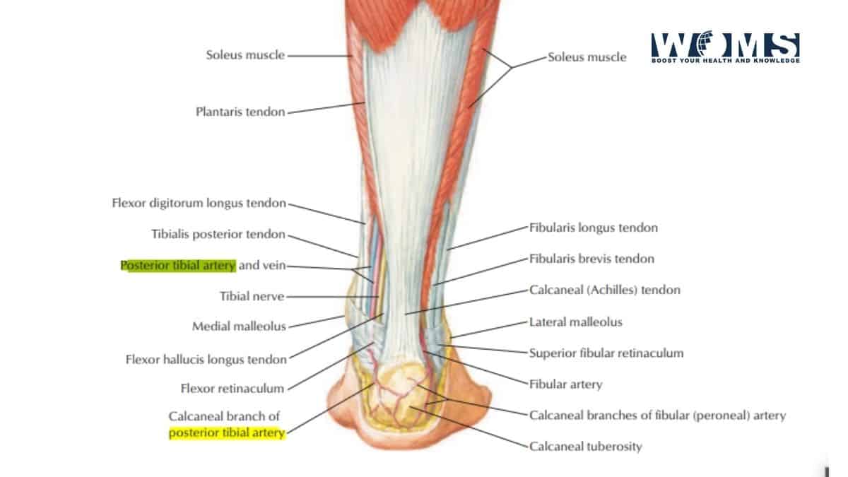
Now that the artery has entered the human leg, it traverses a path directed downwards and medially and reaches the ankle, between the medial tubercle of the calcaneum and the medial malleolus. It ends the route beneath the flexor retinaculum, a thin tendinous sheath, and the origin of the abductor halluces muscles by supplying two terminal branches, the lateral and medial plantar arteries.
Relations of Posterior Tibial artery
During its decent, the tibial nerve and veins of the posterior compartment accompany the posterior tibial artery. The artery courses along the posterior surface of the medial malleolus. The tendons of tibioperoneal trunk and flexor digitorum longus muscle separate the artery from the medial malleolus. Inferiorly, the tendons of the flexor halluces longus and flexor digitorum longus muscles are present on its sides.
Superficial relations of posterior tibial artery:
In the upper two-thirds of the leg, it lies deep to the gastrocnemius, the soleus, and the superficial transverse fascial septum. In the lower part of the leg, it runs parallel to and anterior to the medial side of the Tendocalcaneus. At the ankle, it runs beneath the flexor retinaculum, and the abductor halluces muscles.
Deep relations of posterior tibial artery:
In the upper two-thirds of the leg, it runs along the tibialis posterior. In the lower one-third of the leg, it lies on the flexor digitorum longus and on the tibia. At the ankle, it is placed directly on the capsule of the ankle joint between the flexor digitorum longus and the flexor halluces longus muscles.
Blood supply
It supplies blood to the structures present in the posterior compartment of the leg which include:
- Superficial muscles group: gastrocnemius, soleus and plantaris
- Deep muscles group: flexor Digitorum Longus, flexor halluces Longus and tibialis posterior
- Fasciae: Superficial fascia of the back of the leg, interosseous membrane
- Anastomosis: anastomosis around the ankle, anastomosis around the knee
Size Pecularities
The posterior tibial artery is smaller in size in many people an in those persons it is replaced by the large peroneal artery which continues till the sole of the foot and supplies structure all along its way.
Parent artery, The popliteal artery
The femoral artery gives off the popliteal artery when the femoral artery passes through the adductor hiatus or adductor Magnus. The popliteal artery passes downward and laterally to reach the lower border of the popliteus muscle. The popliteal artery courses along with the joint capsule of the knee. you can also palpate the popliteal pulse behind the knee.
Branches
The posterior tibial artery supplies two terminal branches medial and lateral plantar arteries which supply the sole of the foot. It gives off eight branches in total.
1. Fibula or peroneal artery
It is the largest branch of the tibial artery, arises below the lowermost border of the popliteus muscle and the tendinous arch of the soleus. It runs towards the fibula in an oblique fashion and descends along the medial end of fibula. The nerve to the flexor halluces longus accompanies it along its course through the fibula.
Branches of fibular artery:
The fibular artery gives muscular branches which supply blood to the muscles in both the posterior and the lateral compartments of the human leg.
It also gives rise to the nutrient artery of the fibula.
The fibular artery gives inflation to a perforating branch.
The lateral calcaneal branches supply the heel and join the lateral malleolar network byjoining other malleolar branches in the anastomosis of the ankle.
It also gives off a communicating branch which anastomosis with the similar branch from the posterior tibial artery, above the lower end of the tibia.
2. Circumflex fibular artery
It can arise from either the anterior or the posterior tibial artery at the knee. It runs along the lateral side of the fibula and finally joins the anastomosis around the knee.
3. The nutrient artery of tibia
It is the greater nutrient artery in the human body and It, too, can arise from either the anterior or the posterior tibial artery. The nutrient artery of the tibia supplies blood to the tibialis posterior by piercing it. It enters the nutrient foramen of the tibia which is present in the proximal 3rd of the posterior surface of the bone.
4. Medial plantar artery
The medial plantar artery is the lower terminus branch of the posterior tibial artery. It supplies blood mainly to the great toe by giving off a deep branch. The branch of posterior tibial artery called the medial plantar artery which is an important terminal branch supplies an angiosome via its superficial and deep branches.
Branches of the medial plantar artery:
The trivial branch of the medial plantar artery provides blood to the skin of the medial side of the sole. The superficial branch of the medial plantar artery joins the superficial plantar arch by anastomosing with the lateral plantar artery.
It gives off a deep branch which supplies blood to the great toe and forms three superficial digital branches.
It also gives off many digital branches that associate with digital branches of the medial plantar nerve.
5. Lateral plantar artery
The lateral plantar arteryarises from the posterior tibial artery and the lateral plantar nerve accompanies it. It courses beneath the adductor hallucis and then passes in the space amid the flexor digitorum brevis and quadratus plantae muscles.
Branches of lateral plantar artery:
It gives off muscular branches which supply the adjoining muscles.
The lateral plantar artery also gives off many cutaneous branches that supply blood to the skin and fasciae of the lateral part of the sole of foot.
Anastomotic branches which anastomose with the arteries present on the dorsum of the foot.
A calcanean branch is given off occasionally to the skin of the heel.
6. Muscular branches
Muscular branches of the posterior tibial artery serve as a source of blood supply to the muscles in the posterior compartment of the human leg namely, the soleus, gastrocnemius and plantaris muscles.
- Anastomotic branches
The anastomotic branches join the other arteries around the ankle joint.
- Venae comitantes
Venae Comitantes of the posterior tibial artery joins those of the anterior tibial artery in the popliteal fossa to form the popliteal vein.
Clinical Correlates
1. Posterior tibial pulse:
The posterior tibial pulse can usually be felt behind the medial malleolus and between the flexor digitorum longus and flexor halluces longus tendons.
Ask the person to invert his foot so that the flexor retinaculum is relaxed, and the posterior tibial artery pulse can be easily palpated. It the foot is not inverted and the retinaculum is not relaxed, it can lead to thefalse perception that a pulse is absent.
The posterior tibial pulse is absent is 15% of population. It is an important measure in examining patients with occlusive peripheral arterial disease.
Absence of posterior tibial pulse can hint towards occlusive peripheral arterial disease in patients older than 60 years. For example, the posterior tibial pulse is palpated in cases of intermittent claudication. Intermittent claudication is a disease characterized by severe leg muscle cramps and severe pain. The cramps and severe pain can be a result of lack of blood supply to the leg muscles caused by occlusion of the arteries in the leg.
2. Arterial catheterization
The posterior tibial artery is an alternative site for arterial catheterization. For heart patients who require a number of catheterizations, surgeons usually spare the femoral artery and use the posterior tibial artery for catheterization if possible. However, the posterior tibial artery is smaller and it is relatively difficult to cannulate and can cause an increased risk of thrombosis or ischemia.
Catheterization of this artery is not performed in cases of diabetes mellitus and peripheral vascular disease to save from any complications.
3. The posterior tibial artery perforator flap
The posterior tibial artery perforator flap is used to reconstruct any site of skin loss or injury. In case of loss of soft tissue, the reconstruction requires replacing muscle along with the skin flap. It is generally used in cases of chronic osteomyelitis. The other forms used are:
- Flow-through flap
- Chimeric flap
- Extended flap
- Fascial flap
- Long island flap