What is The Synovial membrane?
Introduction
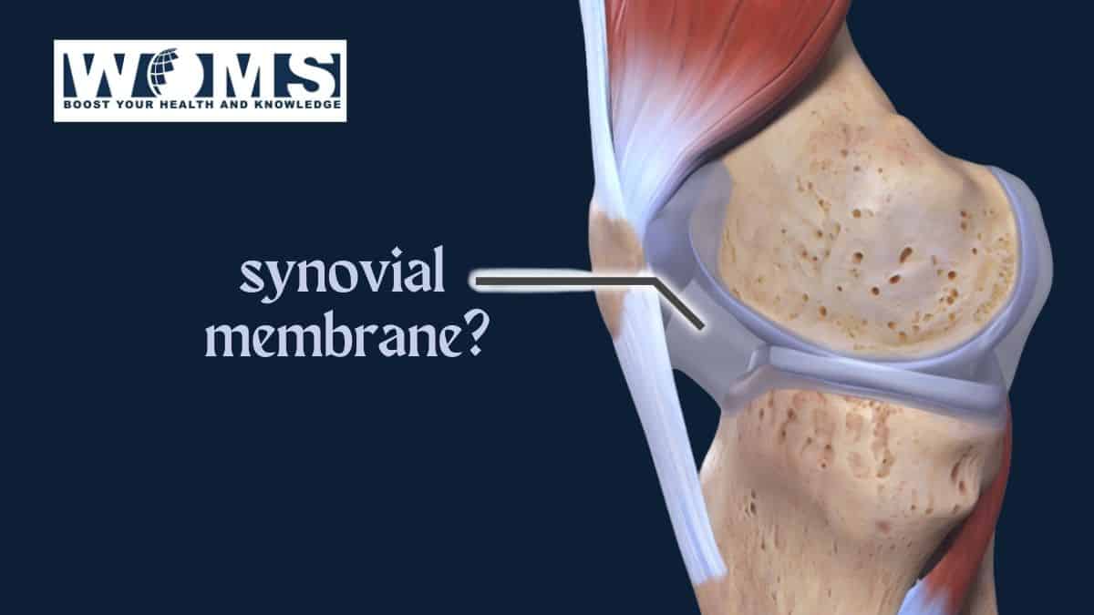
The synovial membrane is a layer of specialized connective tissue. It can simply be explained as a fluid-filled sac in-between joints. And this sac is filled with a fluid called synovial fluid. The membrane lines the inner surface of the fibrous joint capsule of synovial joints. Further, it also lines many other structures inside the joint capsule. Namely, they are
- Exposed osseous surfaces.
- Intracapsular ligaments.
- Bursae.
- Tendon sheaths.
However, it doesn’t cover inter-articular discus or menisci. Therefore, stops at the margins of articular cartilages.
Structure of synovial membrane
Important structures that are made of the membrane
The synovial membrane has few small villi on its internal surface. These villi increase in size and number with age. Some folds and fringes may also protrude into the joint cavity. Examples are the alar fold and ligamentum mucosum of the knee joint. Surfaces of these folds and fringes are abundant with synovial villi. These become prominent in some pathological conditions.
Another important structures are the articular fat pads. Which are formed by the accumulation of adipose tissue within the synovial membrane. Fat pads and folds and fringes take a potential space within the joint. But these spaces are irregular. Because the above structures are not fully filled by synovial fluid. Therefore, they can act as deformable cushions.
Functions of these structures are namely
- Accommodating movement by changing shape and size. By acting alone with intraarticular discs and menisci.
- Increase the area of the synovial membrane.
- Therefore, helping to spread synovial fluid over the articular surfaces.
Layers of the synovial membrane
The synovial membrane has two layers. The outer layer is the subintimal layer. And the inner layer is the intimal layer. The subintimal layer (sub-synovial tissue) is fibrous and vascular. In contrast, the intimal layer is highly vascular.
The subintimal layer can consist of different types of connective tissues. But mostly it is the loose, irregular connective areolar) tissue. Other than that it can also have organized collagen and elastin fibers (dense connective tissue) and adipose tissue. Fibers in dense connective tissue lie parallel to the synovial membrane surface. Elastic fibers prevent the formation of unnecessary folds during joint movement. There are also different types of cells in it. Namely, occasional fibroblast, macrophages, mast cells, and fat cells. Adipose cells form compact lobules. Fibro-elastic interlobular septae surround the adipose lobules. These provide firmness and compressive turgor to the synovial membrane.
The intimal layer is in contact with the synovial fluid. It has synovial cells. These are arranged as very thin sheets of paper. Synovial cells are pleomorphic. The matrix of this tissue is granular and amorphous. Their synovial cells form interlacing discontinuous layers. The thickness of 1-3 cell layers. These cell layers are not separated from the sub intima by a basal lamina.
Cells of the synovial membrane
Synovial cells are elliptical. They have processes that interdigitate. Therefore, they lie close to each other. But gaps separate them from each other. These cells do not normally divide in the synovial membrane. But they will divide in response to trauma and haemarthrosis.
Types of synovial cells.
There are two types of cells in the synovial membrane. They are also called synoviocytes. Namely, they are
- Type A cells.
- Type B cells.
The Type A cells-
These cells are like macrophages. Monocytes in blood give rise to these cells. Accounts for 25% of cells lining the synovium. It has many characteristic organelles. Examples are,
- the surface ruffles or lamellipodia,
- plasma membrane invaginations
- associated pinocytic vesicles
- prominent Golgi apparatus
- little but rough endoplasmic reticulum.
The main function of these cells is the removal of joint debris. Phagocytes do this. And by the synthesis and release of lytic enzymes. Golgi apparatus is prominent for this function of the cells.
Type B cells-
These cells are like fibroblasts. Mesenchyme gives rise to these cells. Its component organelles are different from type A cells. The most important thing is the predominant rough endoplasmic reticulum. They have few vacuoles and vesicles. And less raffled membrane than type A cells. They have many functions.
Functions of Type B cells are namely
- Synthesis of some hyaluronan of synovial fluid. It is a long-chain sugar polymer. They don’t secrete the water in the synovial fluid. But hyaluronan effectively traps water in the joint space.
- Synthesize boundary lubricant lubricin. This is a specialized lubricating protein. It lubricates the joint surface.
These two get together and make the synovial fluid “ropy” like egg white.
- Inhibit the degradative enzyme synthesis by type A cells. This limits their potential to damaged joint tissue.
As previously said during trauma and haemarthrosis these cell types divide. Then type B cells divide in situ. But type A cells increase by immigration of bone marrow-derived precursors.
Synovial fluid and its importance
The synovial membrane secretes and absorbs a fluid called synovial fluid. This fluid fills the inside of the joint capsule. The synovial fluid reduces friction between joints during movements. The fluid acts as a lubricant and does this. This fluid occupies synovial joints, bursae, and tendon sheaths. Its color is pale yellow. And it is a viscous fluid. Inside the synovial membrane, it is slightly alkaline at rest. But during activity, the ph might lower. It contains mixed cells and metachromatic amorphous particles. The fluid is not voluminous. Usually, it is less than 0.5 ml.
Synovial fluid is a transudate of blood plasma. Which is the ultra-filtrate of plasma. Therefore, its composition is consistent. And it is very much similar to plasma. It contains proteins derived from blood. Hyaluronan determines the viscoelasticity of the synovial fluid. It also determines the thixotropic properties. Thixotropic property is the flow rate dependency.
Only a very small proportion of these proteins differ from blood proteins. To be exact around 2%. And that 2% is mostly derived from type B cells. Previously mentioned lubricin is present in an even smaller proportion. Approximately 0.5%. Synovial fluid contains very few cells. In resting cells, it is around sixty cells per millimeter. These cells include monocytes, macrophages, synovial intimal cells, and polymorphonuclear leukocytes. A higher number of cells are present in younger people. During wear and tear, synovial fluid produces by-products. There are two such by-products. Namely, the previously mentioned metachromatic amorphous particles and fragments of cells and fibrous tissue.
Functions of synovial fluid.
- Lubricate the joint.
- Absorbs shock.
- Nourishes the joint capsule.
- Allows smooth movement of joint.
- Reduces friction between articular surfaces.
- Remove debris during wear and tear.
Blood supply and innervation of the synovial membrane.
However, the blood supply to the synovial joints is via articular arteries.
They arise from the vessels around the joint. These are the anastomosing arteries. Mostly the synovial membrane carries these articular arteries.
The nerve endings in the synovial membrane exclusively supply blood vessels. Dues to this it is assumed that the synovial membrane is normally relatively insensitive to pain. But the synovial joints are innervated. There is a law called Hilton’s Law. “The nerves supplying muscles which moves the joint also supplies jointly”. Some of these nerves go to fibrous capsules. Others innervate the capsule and reach the synovial membrane.
Synovial joints
Synovial membranes are seen in synovial joints. These joints are also called diarthrosis. These joints have a fibrous capsule. The fibrous capsule continues with the periosteum of the joined bones. The synovial membrane lines the capsule from the inside. Therefore, the joint capsule can be distinguished into two layers. The outer layer is the fibrous articular capsule. it keeps the bones together. The inner layer is the synovial membrane. It seals in the synovial fluid.
Typically, synovial joints can move fully and freely. They are the most movable joints in the human body. Also the commonest joint type in the body. But they also provide stability in some planes. Intrinsic stability provided by the articular surface and ligament is important.
Mechanisms of the synovial membrane inside the joint.
The articular surface is with low friction due to the synovial membrane. This facilitates the smooth movement of the joint. During rapid movements, microscopic projections on the cartilage surfaces trap synovial fluid. Only small amounts are trapped like this. a thing called fluid film lubrication occurs by this. This is also called akin to aquaplaning. This mechanism reduces friction greatly.
Another feature assists this fluid film mechanism. It is the joint incongruity. Meaning that the opposite articular surface has slightly different curvatures. This produces a potential fluid-filled gap. This gap moves as the joint moves. This helps to wash the synovial fluid across the cartilage surface. This incongruity also reduces peak loading on the apex of the joint.
The synovial fluid can stay longer than water in-between cartilage surfaces. This is because of its sticky and viscous nature. The joint would squeeze out the water too quickly.
Which joints have synovial membranes?
There are seven types of joints that have synovial membranes in the human body. Which are the synovial joints as mentioned above.
- Plane joints.
- Hinge joints.
- Pivot joints.
- Condyloid joints.
- Saddle joints.
- Ball and socket joints.
- Compound joints.
| Type of joint. | Examples. |
| Plane joint/ Gliding joint. | Wrist- in between carpal bones. Shoulder- acromioclavicular joint. |
| Hinge joint. | Elbow- in-between humorous and the ulnar. Knee joint. |
| Pivot joint/ rotary joint. | Joint between ulnar and radius- both distal and proximal. Joint between first and second vertebrae. |
| Condyloid joints/ ellipsoid/ gliding joint. | Joint in the wrist. |
| Saddle joint. | The base of the thumb- carpometacarpal joint. The base of neck- sternoclavicular joint. |
| Ball and socket joint. | Shoulder joint. Hip joint. |
| Compound joints/ bicondyloid joints. | Knee joint- in-between femur, tibia and patella. |
The main disease encountered in the synovial membrane is synovitis. Which is the inflammation of the synovial membrane. Synovitis is when the synovial membrane becomes inflamed. In this, the synovium becomes irritated and swollen. The thickness of the membrane increase.
Causes for synovitis
Injuries, osteoarthritis, and inflammatory arthritis types cause synovitis.
- Overuse of the joint is the most common cause in a healthy person.
Particular occupations are at high risk of developing this. These occupations involve repetitive stress on the joints. For example, weight lifters, athletes, and housewives.
2. Pathologically synovitis is seen in the forms of inflammatory arthritis.
In this the synovium grows excessively. This is happening due to an autoimmune response. In this, the body identifies cartilages as foreign bodies and attacks them. Therefore, in the end, all these damage articular cartilages.
Pathogenesis of synovial membrane in inflammatory arthritis.
One of the most common examples is rheumatoid arthritis. Rheumatoid arthritis is a chronic inflammation of autoimmune origin. It principally attacks the joint. This produces non-suppurative proliferative and inflammatory synovitis. This often leads to the destruction of the cartilage.
In the pathogenesis of rheumatoid arthritis Synovial cell, hyperplasia and proliferation are characteristic. Type B synovial cells play a major role in this. Aggressive forms of these cells are seen. These cells proliferate in-situ.
Meanwhile, an influx of inflammatory cells is seen. pre-existing type A synovial cells also play a role in this. Other than that macrophages also recruit from the outside. An influx of lymphocytes, monocytes, and plasma cells also occurs. Neutrophils and aggregates of organizing fibrin are seen on the synovial surface. With these changes thickness of the synovial membrane is increased. This restricts the physical movement of the joint.
Type B cells produce less amount of hyaluronan. Therefore, lubrication f the cartilage surface is reduced. Synovial cells also produce proteinases. They digest the cartilage matrix. These digested fragments also irritate the synovium. This worsens the inflammatory reaction.
Symptoms of synovitis.
- Joint pain. (arthralgia). The appearance of the joint and the severity of the pain are controversial. Although the joint looks intact pain can be very severe.
- Some systemic symptoms like myalgia, fatigue and generalized musculoskeletal pain were seen.
- Joint stiffness and limitation of movements can be seen.
- Involved joints are swollen, warm and painful.
All these symptoms are for short time. And felt at different parts of the body at different times. But the pain in overuse synovitis is in one place.
Investigations for synovitis.
MRI or musculoskeletal ultrasound are investigations of choice. The main aim is to exclude differential diagnosis from true synovitis. For example, tendonitis can mimic synovitis.
Treatment of synovitis.
- Non-pharmacological- Enough rest.
- Pharmacological- anti-inflammatory medications.
- DMARDS – These are disease-modifying anti rheumatoid drugs. These are mostly immunosuppressing drugs. Examples are Methotrexate and TNF antagonists.
- Corticosteroids.
3. Surgical – if the patient doesn’t respond to these treatments surgery is applicable. This is called a synovectomy. In this, a surgeon removes the synovial membrane. This could be a part of it or fully.
FAQs:
1. How to distinguish between A and B synoviocytes?
When we take morphologically they are macrophages and fibroblasts. We can’t distinguish which one is from the intimal layer. Nor which one is from the subintimal layer. A and B synovitis differ mainly due to their place of existence in the synovial membrane. So they can’t be distinguished separately. We can only say if they are fibroblasts or macrophages.
2. What fluid has the potential to replace the human synovial fluid.
The specialty of synovial fluid is that it contains a higher amount of hyaluronate. In the treatment of osteoarthritis hyaluronic acid can alone be injected into joints. But so far there aren’t any fluids found that can replace synovial fluid fully.
3. Is open surgery synovectomy in RA still popular in comparison with old days.
Drug therapies now are undoubtedly efficient than back then. But there are many patients who don’t respond to drug treatments. Then we have to consider surgery.
4. When should a synovial biopsy be done?
It is not a routinely done test. It is done we can’t come to a conclusion with normal routine investigations. When there’s a suspicion of possible malignancy this can be done.
5. Why does synovial fluid become less viscous.
During inflammations amount of hyaluronic acid produced reduces. This leads to reduced viscosity.